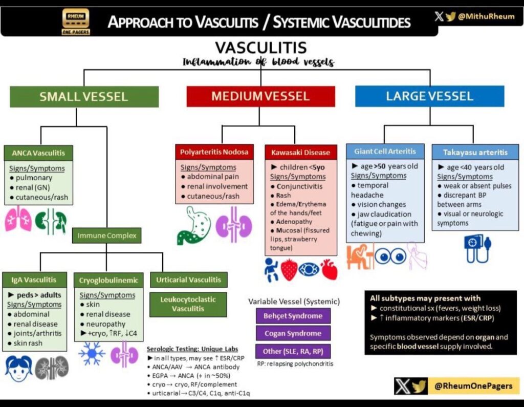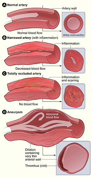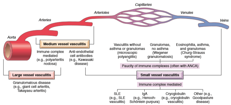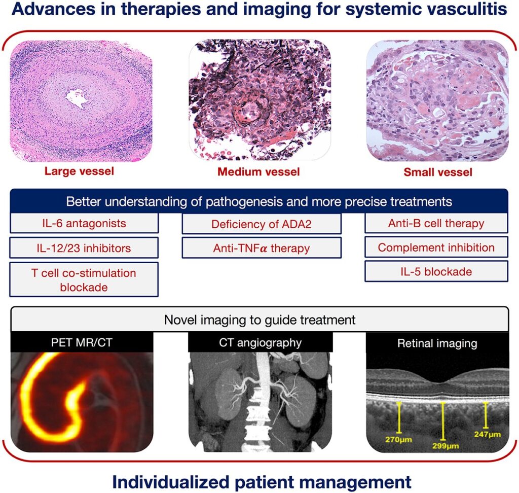
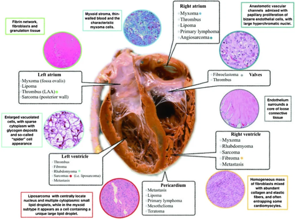
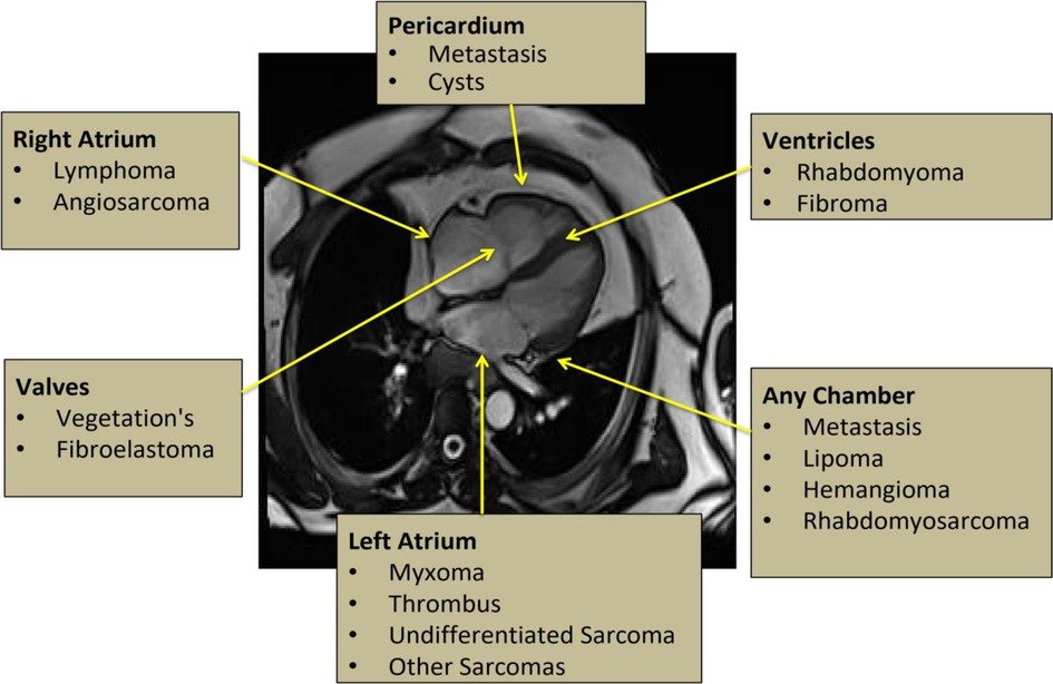
https://www.frontiersin.org/journals/cardiovascular-medicine/articles/10.3389/fcvm.2023.1009411/full
https://link.springer.com/article/10.1007/s11886-020-01420-z
https://www.ahajournals.org/doi/10.1161/CIRCIMAGING.123.016115
Bu necə olur? Bu həqiqətən də belə ola bilər? Yoxsa sadəcə bir hissiyyatdır? Ürək vurğularının ritmini necə bilmək olar? Bunu Həkimə getmədən tayin etmək olarmi?
Dediklərimizi dəqiqləşdirmək üçün ilk növbədə Ürək vurğuları haqda qısa məlumat verək.

Normal halda Ürək 4 kameradan ibarət olur. İki qulaqcıq və iki mədəcik. Qan venalar vasitəsilə əvvəl qulacıqlara daxil olur. Sonra isə İkitaylı və Üçtaylı qapaqlardan keçərək mədəciklərə daxil olur. Buradan isə qanın venoz hissəsi (oksigenlə zəif olan) Ağciyərə digər arterial hissəsi (oksigenlə zəngin olan) isə bütün bədən orqanlarına yayılır.Bu zaman növbə ilə əvvəl ürəyin qulaqcıqları sonra isə mədəcikləri yığılır. Bütün proses təqribən 0,43-0,44 saniyə davam edir. Sonra Ürək 0.40 saniyə dincəlir.Sağlam insanlarda Ürək təqribən dəqiqədə 60-90 vurğu olaraq ritmik yığılmaqdadır. Ritmik dedikdə, yəni Vurğuların müəyyən ardıcıllıqla davam etməsi nəzərdə tututlur. Ritmlərin Ardıcıllığı və sayı pozulduğu zaman bu hal Aritmiya adlandırılır. Bəzən heç bir xəstəliyi olmayan insanlardada bu ardıcıllıq və say dəyişə bilər. Amma ilk əvvəl belə halın xəstəlik olub olmaması Həkim Kardioloq tərəfindən təsdiq olunmalıdır. Bir çox hallarda isə Aritmiyalar təsadüfi müayinələr zamanı aşkar oluna bilər. Ürək ritminin sayının və ardıcıllığının təyin etmək üçün heç də hər zaman Həkimə müraciət etməyə əsas yoxdur. Belə ki, Siz özünüz bilək və ya boyun arteriyasında (damarında) dediklərimizi yoxlaya bilərik. Sadəcə digər əlinizlə nəbzinizin dolğunluğunu və ardıcıllığını yoxlayın. Sonra saata baxaraq ürək döyümələrinizin 1 dəqiqə ərzində neçə dəfə olmasını qeyd edin. Əgər yuxarıda dediyimiz kimi, Vurğu sayı 60-90 arası və ardıcıl ritmik olarsa demək ki, qorxmağa əsas yoxdur. Amma ən yaxın məsafədə EKG müayinəsi oluna bilərsizsə Bu ən uyğun və düzgün yoxlama olardı.
Lakin heç də hər zaman Ritm və Say normal olmaya bilər. Bu zaman artıq Aritmiya haqda danışmaq lazımdır. İlk əvvəl bilmək lazımdır ki, Ürək yığılmaları onun daxili “kiçik elektrik sistemi” ilə tənzimlənir. Bu sistemin bir neçə tərkib hissələri – Sinus düyünü, AV düyün, Hiss dəstəsi və aparıcı yollar var ki, bunların da hər birinin kiçik problemi ciddi Aritmoloji xəstəliklərin yaranmasına səbəb ola bilər.

Ürək ritm pozğunluqlarının bir neçə növü var. Amma tibb sahəsindən kənar olanlar üçün bunu sadəşələşdirib 3 qrupa bölmək olar:
Bütün yuxarıda dediklərimizi yekunlaşdırsaq deyə bilərik ki, Ürək Ritm Pozğunluqları vaxtında aşkar etmək gələcəkdə yarana biləcək ağırlaşmaların aradan qaldırılması üçün cox vacibdir. Bunun üçün Sizdən heç də çox şey tələb olunmur. Sadəcə ən yaxında olan Mütəxəsis Kardioloqa müraciət edib ən sadə Müayinə üsulları ilə diaqnozu təyin etmək olar.
https://pmc.ncbi.nlm.nih.gov/articles/PMC6558967
Published in final edited form as: Clin Gastroenterol Hepatol. 2018 Apr 21;16(8):1189–1196. doi: 10.1016/j.cgh.2018.04.023
Elizabeth K Speliotes *, Maya Balakrishnan ‡, Lawrence S Friedman §, Kathleen E Corey
Dyslipidemia is common in the general population and increases the risk of cardiovascular disease. Treatment of dyslipidemia is effective in decreasing morbidity from cardiovascular disease. Because the liver is the primary source of cholesterol and other lipids in the body, medications for dyslipidemia, such as statins, target genes in the liver. Furthermore, the liver plays a role in the metabolism of many drugs, including those that are used to treat dyslipidemia. It is not surprising, therefore, that many practitioners are hesitant to prescribe medicines to treat dyslipidemia in the setting of liver disease. This update is aimed at summarizing current understanding of the safety of treating patients who have various liver diseases and dyslipidemia with lipid-lowering drugs (Table 1).
Best Practice Advice: Treatment of Dyslipidemia in Liver Disease
| DILI |
| BPA #1 Statins often (3%) cause benign elevations in serum ALT or AST levels and should not be considered contraindicated in patients with liver disease. |
| BPA #2 Liver biochemical tests are recommended before starting a statin but do not need to be checked routinely while statins are taken unless clinically significant side effects develop. |
| BPA #3 Increases in serum ALT or AST levels to >3 times ULN with evidence of cholestasis (bilirubin >2 times ULN) (in the absence of biliary obstruction) after statins are started generally require that the statin be stopped. A work-up for DILI should include testing for the presence of other underlying causes of liver disease or other medications that may have precipitated the reaction besides or in addition to the statin. |
| BPA #4 If DILI or ALF occurs in a patient taking a statin, other statins should be avoided in that patient. |
| BPA #5 DILI and ALF caused by statins are rare (1 in 100,000 and 1 in 1,000,000, respectively), so fear of developing these effects should not be used to justify avoidance of statins when an individual may benefit from them. |
| BPA #6 Statins are contraindicated in patients with ALF because of the patients’ poor prognosis. |
| BPA #7 Other lipid-lowering medications, such as niacin, ezetimibe, or fibrates, may cause DILI, but such instances are exceedingly rare and should not prevent starting these medications in a patient who may benefit from them. |
| NAFLD |
| BPA #8 Although NAFLD and NASH are not considered traditional risk factors for cardiovascular disease, they are associated with dyslipidemia. The 2013 ACC/AHA guidelines should be used to assess cardiovascular risk in patients with NAFLD and to guide the need for lipid-lowering pharmacotherapy. |
| BPA #9 Statins, ezetimibe, omega-3 fatty acids, and fibrates are safe and well tolerated in the setting of NAFLD and NASH. |
| BPA #10 A statin is first-line treatment of elevated serum LDL levels in patients with NAFLD who are deemed to be at increased risk for adverse cardiovascular disease outcomes. Statin therapy is associated with reductions in serum LDL levels and cardiovascular disease prevention in patients with NAFLD. |
| BPA #11 Ezetimibe may also be used for treatment of elevated LDL levels, either as primary therapy in patients who are statin intolerant or in addition to a statin when the statin is insufficient to reduce LDL levels. Ezetimibe is associated with reductions in LDL levels, but its efficacy for cardiovascular disease prevention is unknown. |
| BPA #12 Omega-3 fatty acids and fibrates are indicated for the treatment of isolated hypertriglyceridemia. |
| BPA #13 There is no conclusive evidence that treatment of dyslipidemia with any agent (statin, fibrate, fish oil) improves the histology of NASH or liver-related outcomes. |
| Viral Hepatitis |
| BPA #14 Despite causing a reduction in serum lipid levels, chronic HCV infection is associated with an increased risk of acute myocardial infarction. Serum LDL and total cholesterol levels rebound after spontaneous and treatment-induced viral clearance. Therefore, lipid levels should be monitored after HCV clearance to determine if a patient has a new indication for treatment of dyslipidemia. |
| BPA #15 The impact of chronic HBV infection on serum lipid levels is not well described, but HBV infection may decrease serum triglyceride and HDL levels. |
| BPA#16 Management of patients with HBV or HCV infection and dyslipidemia should be guided by standard recommendations for the treatment of dyslipidemia. |
| BPA #17 Statins are safe to use in patients with either chronic HCV or HBV infection, but attention should be paid to potential interactions between statins and antiviral agents. |
| PBC |
| BPA #18 Dyslipidemia in the form of elevated serum cholesterol and triglyceride levels is common in PBC, does not increase the risk of cardiovascular disease, and does not need to be treated with lipid-lowering agents unless other concomitant cardiovascular risk factors are present. |
| BPA #19 Lipid-lowering agents, such as statins, are not contra-indicated in patients with PBC with compensated liver disease but should not be used in patients with decompensated disease. |
| BPA #20 Second-line treatments for PBC, such as fibrates and OCA, can affect lipid levels. Until more is known about the effect of OCA on cardiovascular disease, OCA in particular should be avoided in patients with PBC who have cardiovascular disease or risk factors for disease. OCA should be dosed weekly rather than daily in PBC patients with Child-Pugh class B or C cirrhosis. |
| BPA #21 There is no compelling evidence that statins can improve outcomes in patients with PBC; they should not be used as primary agents for treatment of this disease. |
| Cirrhosis |
| BPA #22 Statins can be safely used in patients with Child-Pugh class A cirrhosis for cardiovascular risk reduction if indicated. |
| BPA #23 Statins should be avoided in patients with Child-Pugh class B or C cirrhosis because of the patients’ poor prognosis, not because of increased hepatotoxicity. |
| Post-Transplant Dyslipidemia |
| BPA #24 Dyslipidemia is common following liver transplantation, affecting up to 62% of transplant recipients. Pretransplant obesity and diabetes mellitus increase the risk of post-transplant dyslipidemia. Post-transplant weight gain and immunosuppres-sant medications, including calcineurin inhibitors and the mTOR inhibitor sirolimus, also increase the risk of post-transplant dyslipidemia. |
| BPA #25 Lipid-lowering agents, specifically statins, are not associated with an increased risk of hepatotoxicity in the post-transplant population and may be used as needed to treat dyslipidemia. |
| BPA #26 Calcineurin inhibitors, like several statins, are metabolized by CYP3A4 and may increase the risk of statin-associated myopathy. Pravastatin and fluvastatin are not metabolized by CYP3A4 and do not increase the risk of statin-associated myopathy when used with a calcineurin inhibitor. |
The 2013 American College of Cardiology/American Heart Association guidelines, published in 2014, should be used to guide treatment of dyslipidemia in patients with the liver diseases discussed in this update.1 The guidelines recommend that adults with cardiovascular disease or a low-density lipoprotein (LDL) level 190 mg/dL be treated with high-intensity statins, with the goal of reducing LDL levels by 50%. Individuals 45–70 years of age with diabetes mellitus and a serum LDL level <189 mg/dL or persons with a >7.5% global 10-year risk of cardiovascular disease can be treated with moderate-intensity statins, with the goal of reducing LDL levels by 30%–50% (Table 2).
Indications for Pharmacologic Reduction of Serum LDL Levels in the General Population
| Presence of clinical atherosclerotic cardiovascular disease (coronary heart disease, symptomatic carotid artery disease, stroke/transient ischemic attack, peripheral artery disease, abdominal aortic aneurysm) |
| Adults 40–77 years of age with diabetes mellitus and LDL levels of 70–189 mg/dL |
| Adults 40–75 years of age with a global 10-year risk of cardiovascular disease ≥7.5% and an LDL level of 70–189 mg/dL |
| Adults with an LDL level ≥190 mg/dL |
Adapted from 2013 American Heart Association/American College of Cardiology guidelines.1
Drug-Induced Liver Injury
From 8% to 9% of persons in the general population have an elevated aminotransferase level, a common clinical problem.2 In these persons, an evaluation to determine the cause of the aminotransferase elevation is warranted, particularly before a new drug is started, because they may have a common underdiagnosed condition, such as nonalcoholic fatty liver disease (NAFLD), excessive alcohol use, or viral hepatitis. Statins (3-hydroxy-3-methyl-glutaryl-coenzyme A reductase inhibitors) are by far the most common medication used to treat dyslipidemia and are known to cause elevations of serum alanine aminotransferase (ALT) levels in persons with previously normal levels. Overall, persons on low-to-moderate statin doses have, on average, a 1% chance of having an elevated ALT level.3 At most, 3% of patients on statins develop elevations in serum ALT levels.3 The effect is generally dose dependent, with higher doses of statins increasing the chances of an elevated ALT level. These mild elevations do not generally indicate serious toxicity.
Elevations in aminotransferase level indicative of serious liver injury caused by a drug are rare but can be life threatening. Zimmermann4,5 observed that jaundiced patients with elevated aminotransferase levels had a poorer prognosis than patients with elevated amino-transferase levels without jaundice. That observation was further developed into Hy’s Law to identify patients with potentially fatal drug-induced liver injury (DILI).6 To meet criteria for DILI, patients must and have elevations of ALT or aspartate aminotransferase levels ≥3 times the upper limit of normal (ULN) and have elevations of the total serum bilirubin level >2 times the ULN with no other identified cause of the increased liver biochemical tests (eg, biliary obstruction, another liver disease, other drug toxicity) except for the offending drug.6 DILI from statins occurs in 1 in 100,000 persons and can have a variety of histologic presentations.7,8 There can be an asymptomatic rise in the ALT level (<3 times ULN) that can improve with continued stain use, so-called adaptation; hepatitis with an ALT level >3 times ULN and clinical liver disease; cholestatic hepatitis with development of jaundice; and autoantibody-associated DILI with the development of positive antinuclear antibodies and antimitochondrial or smooth muscle antibodies with or without plasma cells in liver biopsy specimens. In general, however, statins can be used in individuals with autoimmune disorders including autoimmune hepatitis. General care should be taken to ensure that medications taken for autoimmune hepatitis or other disorders do not affect the catabolism or excretion of statins, thereby altering their effective dose. Statins can also cause acute liver failure in 1 in 1,000,000 persons.
The liver safety of statins has recently been reviewed and guidelines published for their use.9 It is recommended that liver biochemical tests be checked before a statin is started. However, routine periodic monitoring of serum ALT levels does not seem to detect or prevent serious liver injury in association with statin use and is not recommended. Only if and when a patient shows clinical signs, such as fever or jaundice, of adverse effects from a statin should liver biochemical tests be rechecked. If DILI from a statin is suspected, then a full work-up should be initiated to ensure that the cause is the statin and not a secondary liver disease or another drug (reviewed in Ref.9).
Niacin, ezetimibe, and fibrates, which are also used to treat serum lipid disorders, have all been reported to cause DILI. If a patient had DILI or acute liver failure from a statin, this class of medications should not be used in that patient again, and an alternative class of lipid-lowering drugs can be tried.7 Statins are contraindicated in patients with decompensated liver disease or acute liver failure (see later) but are otherwise safe, even at high doses, and efficacious in lowering lipid levels in patients with compensated liver disease.10
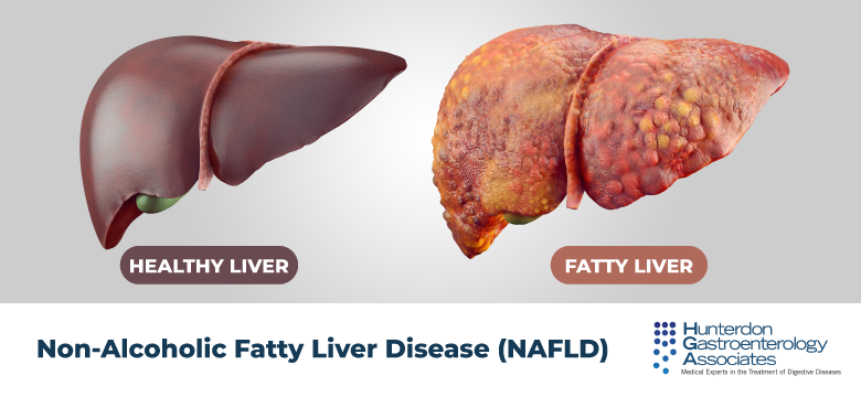
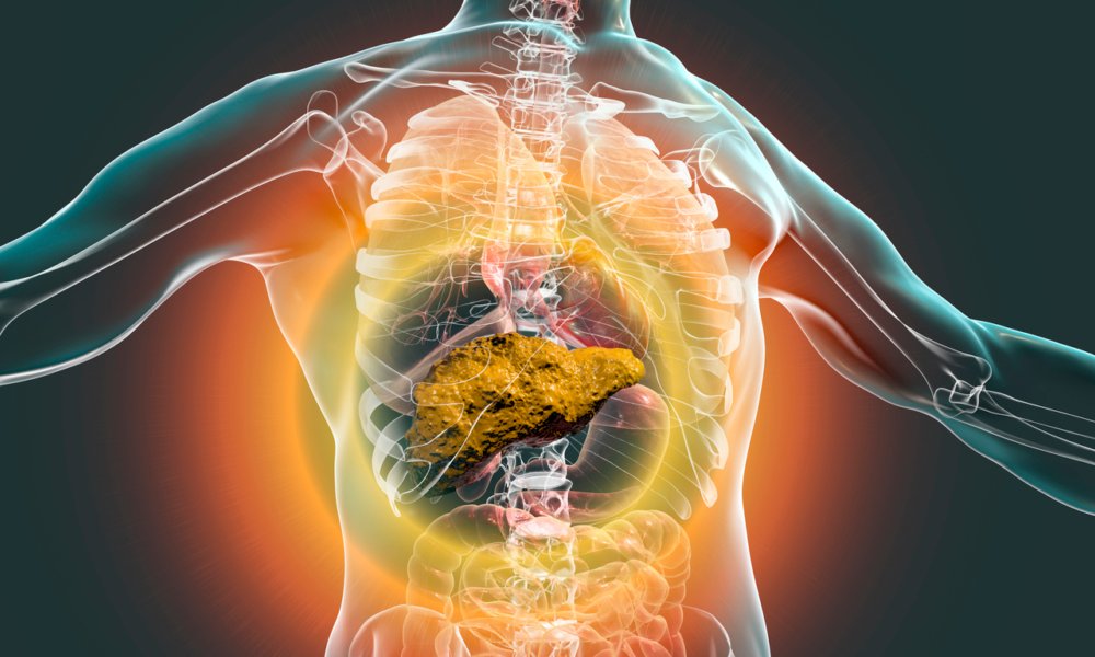
Dyslipidemia is common among persons with NAFLD and plays a critical role in the development of cardiovascular disease, the leading cause of mortality among these persons. Dyslipidemia associated with NAFLD is typically characterized by a pattern of hyper-triglyceridemia, increased LDL levels, and reduced high-density lipoprotein (HDL) levels. Although patients with NAFLD have a higher risk of cardiovascular complications compared with the general population, NAFLD and nonalcoholic steatohepatitis (NASH) are not considered traditional risk factors for cardiovascular disease. Therefore, cardiovascular risk should be assessed according to the 2013 American College of Cardiology/American Heart Association guidelines, which use presence of known coronary artery disease, cardiovascular risk factors, and 10-year cardiovascular event risk assessment to determine the need for treatment with lipid-lowering agents and to establish LDL targets for primary and secondary cardiovascular risk prevention (Table 2; discussed later).
Diet and exercise are the first-line approaches to reduce LDL levels. When diet and exercise are insufficient, statins are the first-line pharmacologic agents for LDL reduction and have proven benefit for primary and secondary prevention of cardiovascular disease. Several studies have demonstrated that statins are effective in reducing LDL levels and cardiovascular events and are safe in patients with NAFLD. Intensive statin therapy (eg, pravastatin, 80 mg daily) was associated with a significant reduction in LDL levels compared with placebo among patients with chronic liver disease primarily caused by NAFLD (mean percentage LDL reduction, 30.6%; standard deviation, 16.5%).10 In a post hoc analysis of the ATTEMPT study, a greater benefit in primary cardiovascular disease prevention was observed among patients with NAFLD and metabolic syndrome in whom LDL reduction was aggressive with intensive statin therapy (atorvastatin titrated from 10 to 80 mg daily) aimed to reduce LDL levels to <100 mg/dL compared with those in whom the LDL reduction was aimed to reduce the level to <130 mg/dL.11 Post hoc analyses of 2 large randomized controlled trials (RCTs) (GREACE and IDEAL studies) also found superior secondary cardiovascular disease prevention benefit in association with intensive statin therapy among patients with increased aminotransferase levels attributed to NAFLD. In the IDEAL study, compared with moderate statin therapy, intensive statin therapy (atorvastatin, 80 mg daily) was associated with a 44% relative risk reduction in secondary cardiovascular events (hazard ratio, 0.556; 95% confidence interval, 0.367–0.842).12 In the GREACE study, compared with usual care, intensive statin therapy (atorvastatin titrated from 10 to 80 mg daily for an LDL goal of <100 mg/dL) was associated with a 68% relative risk reduction (P < .0001).11 Both studies found greater cardiovascular benefit among patients with increased aminotransferase levels than in those with normal levels.
Statins are well tolerated and not associated with liver-related adverse events among patients with NAFLD and dyslipidemia.10,13,14 In RCTs, patients with NAFLD who were treated with intensive statin therapy experienced significant reductions in aminotransferase levels compared with control subjects.10 Observational studies have similarly reported significant reductions in amino-transferase levels and steatosis on liver biopsy specimens in statin-treated patients with NAFLD. However, histologic improvements in ballooning and fibrosis have not been observed in association with statins.
Experience with ezetimibe in patients with NAFLD is much less extensive than that with statins. Treatment with ezetimibe, 10 mg daily, was associated with significant reductions in LDL and total cholesterol levels among patients with NAFLD in several observational studies15–18 and 1 small open-label RCT.19 However, there have been no studies looking at the effect of ezetimibe on cardiovascular disease prevention in patients with NAFLD. Ezetimibe may be associated with improvements in aminotransferase levels. Mixed results from studies evaluating the effect of ezetimibe on liver histology in patients with NASH suggest that ezetimibe may improve steatosis, NAFLD activity score, and hepatocyte ballooning but not fibrosis.15,17,19–23
Omega-3 fatty acids and fenofibrates are safe and effective for the treatment of isolated hyper-triglyceridemia. Neither medication is associated with improvement in liver histology in patients with NAFLD.
In summary, statin therapy is indicated for treatment of increased LDL levels among patients with NAFLD who are at increased risk for adverse outcomes of cardiovascular disease. Statin therapy is associated with reductions in LDL levels and prevention of cardiovascular disease. Ezetimibe may also be used for treatment of increased LDL levels and is associated with reductions in these levels, but its efficacy for cardiovascular disease prevention is unknown. Omega-3 fatty acids and fibrates are indicated for the treatment of hypertriglyceridemia. All of these agents are safe and well tolerated in patients with NAFLD. However, there is no conclusive evidence to date that treatment of dyslipidemia with any agent (statins, fibrates, omega-3 fatty acids) improves histology or liver-related outcomes in patients with NASH.
Hepatitis C virus (HCV) chronically infects up to 170 million individuals globally and can lead to the development of cirrhosis, hepatocellular carcinoma, and the need for liver transplantation.24 HCV replication impacts host lipid metabolism via several mechanisms. HCV virions interacts with circulating lipoproteins to infect hepatocytes via the LDL receptor and interacts with the cell surface markers Niemann-Pick C1-like 1, a receptor for cholesterol resorption, and scavenger receptor class B member 1, which promotes uptake of cholesterol from lipoproteins.25–27 Once inside the hepatocyte, HCV interacts with host cytosolic lipid droplets and diacylglycerol O-acetyltransferase 1 to form the HCV core protein and uses an intermediate of the cholesterol synthesis pathway for replication.27 HCV also uses host lipids for secretion by complexing with apoE-containing very-low-density lipoproteins and HDL.28
These HCV-host interactions affect host circulating lipid levels. Both acute and chronic HCV infections lead to a decrease in serum LDL and total cholesterol levels.29,30 However, the lower lipid levels found in persons with HCV infection do not confer a decreased risk of cardiovascular disease; on the contrary, chronic HCV infection is associated with an increased risk of acute myocardial infarction when compared with matched control subjects.31,32 Furthermore, LDL and total cholesterol levels rebound after spontaneous and treatment-induced viral clearance. Therefore, in patients with HCV infection, lipid levels should be monitored, with a special focus on lipid levels following HCV clearance, to determine if treatment of dyslipidemia is needed. Treatment with statins is safe in individuals with liver disease, including hepatitis C, and should be first-line therapy.33
Hepatitis B virus (HBV) chronically infects more than 240 million individuals worldwide and can lead to end-stage liver disease, hepatocellular carcinoma, and the need for liver transplantation.33,34 Like HCV, HBV interacts with lipid metabolism in the host. HBV uses the peptide Naþ-taurocholate cotransporting polypeptide as a point of viral entry and an aid in the production of hepatitis B surface antigen. The binding of HBV to Na+ taurocholate cotransporting polypeptide impairs bile acid uptake, thereby leading to increased bile acid synthesis and bile acid conversion to cholesterol.35 Hepatitis B surface antigen formation also relies, in part, on the host endoplasmic reticulum lipid bilayer and, once formed, is exported as a lipoprotein particle.36
Expression of lipogenic genes may also be altered by HBV infection. Mouse models of HBV infection demonstrate increased expression of genes for SREBP2, 3-hydroxy-3-methylglutaryl-coenzyme A reductase, the LDL receptor, fatty acid synthase, ATP citrate lyase, peroxisome proliferator-activated receptor-ɣ, and apolipoprotein A1.37,38
Data on host lipid levels during HBV infection in humans are limited. A case-control study found that HBV-infected individuals had lower serum triglyceride and HDL levels than age-matched control subjects. A second retrospective study found that serum HBV DNA levels were inversely correlated with serum triglyceride levels. No relationship between serum HDL and HBV DNA levels was seen.39
As for HCV infection, there is a lack of data to guide lipid management in individuals with HBV infection. Clinicians should adhere to guidelines from the American College of Cardiology/American Heart Association on lipid management (Table 2).1 Statin therapy is safe to use in patients with chronic hepatitis B and should not be withheld.
Primary biliary cholangitis (PBC) is a chronic infiammatory autoimmune cholestatic liver disease associated with dyslipidemia. Individuals with PBC have a complex pattern of dyslipidemia (reviewed in Ref.40). Patients with PBC have increased serum triglyceride levels and variable HDL levels: patients with PBC and stage 1 or 2 fibrosis have increased HDL levels, those with stage 3 fibrosis have variable HDL levels, and those with stage 4 fibrosis have decreased HDL levels. These patients have increased total cholesterol levels largely because of elevations of lipoprotein X. Lipoprotein X is antiatherogenic; therefore, even though patients with PBC have dyslipidemia, the dyslipidemia may not lead to an increase in cardiovascular events. It is estimated, however, that about 10% of patients with PBC have a significant risk of cardiovascular disease and should be treated with medications to reduce that risk. Statins, ezetimibe, and fibrates have all been shown to be safe in patients with PBC.
Fibrates are second-line therapies for the treatment of cholestasis in patients with PBC and also affect serum lipids.41 Fibrates have anticholestatic effects through the activation of peroxisome proliferator-activated receptor and downregulation of bile acid synthesis pathways. Benzafibrate, 400 mg daily, alone or in combination with ursodeoxycholic acid, has been shown to lower serum alkaline phosphatase levels in small studies, but concern remains about ascertainment bias and the significance of biochemical rather than histologic or survival endpoints (reviewed in Ref.41). A large RCT is underway, and until the results are available, these medications may be used off-label in patients in whom the response to ursodeoxycholic acid is inadequate. Fibrates, however, do not significantly reduce the risk of cardiovascular events42 and should not be used for primary prevention of cardiovascular disease in patients with PBC and dyslipidemia.
Obeticholic acid (OCA) is a second-line therapy for the treatment of PBC and also affects serum lipids.41 OCA is a farsenoid-X receptor agonist that directly regulates genes involved in bile acid synthesis, secretion, transport, absorption, and detoxification. The phase 3 PBC OCA International Study of Efficacy (POISE)43 showed a 33%–39% reduction in serum alkaline phosphatase levels in the group receiving 10 mg daily compared with 5% in the group receiving placebo (P < .001) at 12 months of treatment. However, patients taking OCA had an increase in LDL and total cholesterol levels and a decrease in HDL levels, the cardiovascular consequences of which remain to be determined. If cardiovascular risk is indeed increased in patients taking OCA, the medication may need to be coupled with a second medication to lower that risk. Thus, until more data are available, OCA should be avoided in patients with cardiovascular disease or risk factors for cardiovascular disease. Additionally, the recommended initial dose for patients with moderate to severe liver impairment (Child-Pugh B and C) is 5 mg once weekly, rather than the 5 mg daily used for other PBC patients (https://www.fda.gov/downloads/Drugs/DrugSafety/UCM576880.pdf). When daily dosing has been administered inadvertently in Child-Pugh B and C patients, serious liver injury and death have been reported. Furthermore, up to 10% of patients discontinued treatment because of pruritus.
Several studies have now shown that statins can lower LDL levels in patients with PBC who also have increased rates of cardiovascular disease. Atorvastatin, 10 mg daily for 1 year, in early stage PBC reduces total cholesterol, LDL cholesterol, LDL, and triglyceride levels without affecting progression of cholestasis.44 Long-term atorvastatin use also led to reductions in total and LDL cholesterol levels.45 Similar reductions in total cholesterol and LDL cholesterol levels were seen in patients treated with simvastatin, 20 mg daily.46 Although initially considered promising treatments for PBC,46 recent trials have not shown an effect of statins in reducing serum alkaline phosphatase levels.44,45 Studies also show that ezetimibe is safe in patients with PBC.47 Therefore, statins and ezetimibe should be considered in patients with PBC with cardiovascular disease risk factors.
Although cirrhosis was previously thought to protect against atherosclerotic disease, work in recent years has demonstrated that the prevalence of coronary artery disease among patients with cirrhosis may be higher than that in the general population.48–50 Cardiovascular risk varies according to etiology of liver disease and is highest in cirrhosis caused by alcohol, HCV, and NASH51–53; in NASH, the risk is mediated by concomitant risk factors for cardiovascular disease or by the presence of steatosis and insulin resistance. The risk of atherosclerosis is highest in NASH cirrhosis.53 Cholestatic liver diseases, however, do not carry an overall increased risk of atherosclerotic disease.51
Statins are safe and can be used in patients with Child-Pugh class A cirrhosis.9 There are little to no available data regarding the safety and risks of statin use in patients with decompensated cirrhosis. The updated 2014 recommendations of the Liver Expert Panel assembled to address the safety of statins in liver disease9 provides some guidance and advises against statin use among patients with Child-Pugh class B or C cirrhosis. The underlying rationale is that the generally grave liver-related prognosis of patients with Child-Pugh class B or C cirrhosis makes it unlikely that they will benefit from the cardiovascular benefits associated with lipid-lowering therapy. In addition, moderate-to-severe hepatic impairment may result in reduced drug metabolism and consequently abnormally high serum drug levels (although an increased risk of hepatotoxicity has not been demonstrated).
In addition to lipid-lowering effects, statins are associated with reductions in portal pressure and may reduce the risk of decompensation among patients with HBV or HCV cirrhosis.54–56 However, the data are still evolving, and use of statins to reduce portal pressure is not the standard of care. Therefore, statins should not be used for the treatment of portal hypertension.
Dyslipidemia is common following liver transplantation, occurring in up to 62% of transplant recipients.57 Post-transplant dyslipidemia is seen most commonly in patients with obesity or diabetes mellitus pretransplantation but can develop in the absence of these comorbid conditions. Weight gain post-transplantation and use of immunosuppressant medications, including calcineurin inhibitors and the mechanistic target of rapamycin inhibitor sirolimus, also increase the risk of post-transplant dyslipidemia.58
Lipid-lowering agents, specifically statins, are safe in the post-transplant population and should be used as needed to treat dyslipidemia.59 Calcineurin inhibitors and several statins are metabolized by cytochrome P-450 3A4, and concurrent use may increase the risk of statin-associated myopathy. Pravastatin and fiuvastatin are not metabolized by cytochrome P-450 3A4 and, when used with cyclosporine, may not increase the risk of statin-associated myopathy.59,60
Lipid-lowering medications are safe and effective in lowering the risk of cardiovascular disease in individuals with compensated liver disease. Some lipid-lowering agents may help in the primary or secondary treatment of specific types of liver disease. Interactions with other drugs should be considered when choosing particular lipid-lowering medications. Only in patients with decompensated cirrhosis and in those with well-documented DILI from a lipid-lowering agent should these medications not be used.
— Recent developments of interest in cardiovascular medicine
by Nicole Lou, Senior Staff Writer, MedPage TodayOctober 29, 2024
A Chinese study suggested that cold weather may make people more susceptible to myocardial infarction with non-obstructive coronary arteriesopens in a new tab or window compared with myocardial infarction from obstructive disease. (European Heart Journal)
Pneumonia and urinary tract infection may act as triggersopens in a new tab or window of acute myocardial infarction, according to a Danish cohort study. (European Journal of Preventive Cardiology)
People with bipolar disorder and schizophrenia had outsized risks of sudden cardiac deathopens in a new tab or window, another Danish study showed. (Heart)
For patients with systolic heart failure and moderate aortic stenosis, preemptive transcatheter aortic valve replacementopens in a new tab or window (TAVR) did not improve clinical outcomes over standard therapy in a small randomized trial. (Journal of the American College of Cardiology)
Early aortic valve intervention also failed to show clinical benefit in those with asymptomatic severe aortic stenosis and myocardial fibrosisopens in a new tab or window in a separate trial. (JAMA)
Cardiawave said its Valvosoft Non-Invasive Ultrasound Therapyopens in a new tab or window device met expectations for 6-month results in a pivotal trial of severe symptomatic calcific aortic stenosis.
A microaxial flow pumpopens in a new tab or window reduced the need for pharmacological circulatory support while maintaining hemodynamic stability in patients with ST-elevation myocardial infarction-induced cardiogenic shock, per a DanGer Shock substudy. (JAMA Cardiology)
Medtronic announced FDA approval of its Affera Mapping and Ablation System with the Sphere-9 Catheteropens in a new tab or window for radiofrequency and pulsed field ablation in persistent atrial fibrillation.
The PRECISE-HBR scoreopens in a new tab or window, which predicts post-percutaneous coronary intervention bleeding, offered a slight improvement in discrimination over existing scores. (Circulation)
Routine upfront use of orbital atherectomyopens in a new tab or window for severely (but not extremely) calcified lesions did not improve stent expansion or patient outcomes compared with routine balloon angioplasty prior to stenting, according to ECLIPSE trial results highlighted by Mount Sinai.
In a head-to-head comparison of two-stent strategies for coronary bifurcation lesions outside the left main, the double-kissing culotte techniqueopens in a new tab or window came out on top. (Circulation: Cardiovascular Interventions)

The sutureless, fully absorbable Vivasure PerQseal Closure Device Systemopens in a new tab or window performed well for large-bore vessel punctures in the PATCH IDE pivotal study, the device maker announced.
Male gender expressivityopens in a new tab or window, reflecting cultural pressures to convey masculinity, in young men was associated with fewer hypertension and diabetes diagnoses and treatment in adulthood, a cohort study suggested. (JAMA Network Open)
Two years after radiofrequency renal denervation with the Symplicity Spyral systemopens in a new tab or window, patients continued to show significant blood pressure reductions in the SPYRAL HTN-ON MED study, Medtronic said.
A first-in-human study supported an investigational cerebroprotection agent — a small-molecule adenosine A1/A3 receptor agonistopens in a new tab or window — for phase II study in stroke and traumatic brain injury. (Stroke)
verfasst von: PD Dr. Antje Schmidt-Pogoda, Frederike A. Straeten, Carolin Beuker, Nils Werring, Jens Minnerup
Erschienen in: Der Nervenarzt | Ausgabe 10/2024
Der ischämische Schlaganfall ist weltweit eine der häufigsten Todesursachen und häufigste Ursache für eine anhaltende Behinderung im Erwachsenenalter. Die weit überwiegende Anzahl von Schlaganfällen ist Folge vaskulärer Risikofaktoren, insbesondere der arteriellen Hypertonie. Die häufigsten unmittelbaren Ursachen des zerebralen Gefäßverschlusses sind hierbei das Vorhofflimmern sowie arterielle Embolien bzw. lokale Verschlüsse infolge atherosklerotischer Gefäßstenosen. Neben diesen häufigen Schlaganfallursachen, die überwiegend zum Erkrankungsgipfel im höheren Lebensalter beitragen, gibt es eine Vielzahl seltener Schlaganfallursachen mit z. T. einer Häufung bei jüngeren Menschen. In diese Kategorie der seltenen Schlaganfallursachen fallen verschiedenste entzündliche systemische wie auch auf das Zentralnervensystem (ZNS) fokussierte Erkrankungen. Sie sind Gegenstand des vorliegenden Übersichtsartikels. Hierbei liegt der Fokus auf den alltagsrelevanten Aspekten Diagnostik und Therapie, die häufig spezifisch sind.
Die primäre Angiitis des zentralen Nervensystems (PACNS) ist eine Vaskulitis unbekannter Ursache, die isoliert die Arterien (und weniger häufig die Venen) des Gehirns, des Rückenmarks und der Leptomeningen betrifft. Die geschätzte Inzidenz der PACNS beträgt 2,4 Fälle pro 1.000.000 Personen pro Jahr, dabei sind Männer und Frauen gleich häufig betroffen. In einer rezenten Analyse von 911 PatientInnen mit PACNS lag das durchschnittliche Patientenalter bei Diagnosestellung bei 42 Jahren, die Altersspanne reichte von 24 bis 63 Jahren. Die definitive Diagnose PACNS kann nur bioptisch oder autoptisch gestellt werden
Da die Vaskulitis jegliche Region des ZNS beeinträchtigen kann, variieren die klinischen Erscheinungsformen. Zu den häufigsten Symptomen gehören fokal-neurologische Defizite (63 %), Kopfschmerzen (51 %) und kognitive Beeinträchtigungen (41 %) Die Kopfschmerzen infolge PACNS werden unterschiedlich beschrieben, neigen jedoch dazu, eher subakut und unterschwellig zu sein, im Unterschied zu den abrupt auftretenden, intensiven Donnerschlagkopfschmerzen, die typisch für das reversible zerebrale Vasokonstriktionssyndrom (RCVS) sind. Schlaganfälle und transiente ischämische Attacken (TIAs) treten oft mehrzeitig und in unterschiedlichen Gefäßterritorien auf.
Die Diagnosestellung der PACNS gestaltet sich oft als herausfordernd. Aufgrund der risikobehafteten Therapieoptionen ist jedoch eine große therapeutische Sicherheit gefordert. Die definitive Diagnose kann nur bioptisch oder autoptisch gestellt werden. Sofern die Diagnose nicht bioptisch gesichert werden kann, lässt sich eine wahrscheinliche PACNS diagnostizieren, wenn sowohl angiographische als auch magnetresonanztomographische (MRT-)Untersuchungen typische Merkmale aufweisen und diese durch ein für die PACNS spezifisches Liquorprofil ergänzt werden (s. unten). Die Entnahme einer leptomeningealen und parenchymatösen Biopsie sollte möglichst aus einem MR-tomographisch oder angiographisch betroffenen Bereich erfolgen. Hierdurch kann die Sensitivität der Biopsie auf bis zu 80 % angehoben werden. Bevorzugte Biopsielokalisation sind nichteloquente Areale der nichtdominanten Hemisphäre. Bei positivem Befund finden sich drei unterschiedliche histologische Muster: granulomatös mit multinukleären Zellen (58 %), lymphozytär (28 %) oder nekrotisierend (14 %). Neben der Diagnosesicherung dient die Biopsie außerdem dem Ausschluss von Differenzialdiagnosen, wie primären ZNS-Lymphomen, Tumoren u. a.
Die konventionelle zerebrale digitale Subtraktionsangiographie (DSA) zeigt bei der PACNS mit Befall mittelgroßer bis großer Gefäße (Medium-to-large-vessel-Variante [MV/LV-PACNS]) typischerweise multilokuläre segmentale Stenosierungen. Diese sind zwar charakteristisch, ausgeprägte arteriosklerotische Veränderungen sowie das reversible zerebrale Vasokonstriktionssyndrom können jedoch ein ähnliches Muster zeigen. Bei der Small-vessel-Variante (SV-PACNS) bleibt die DSA unauffällig. In diesen Fällen ist eine histologische Diagnosesicherung durch eine Hirnbiopsie erforderlich. Magnetresonanztomographisch zeigen sich typischerweise multifokale Läsionen in der weißen Substanz, gadoliniumaufnehmende Läsionen und ein leptomeningeales Enhancement. Gradientenechosequenzen können petechiale Hämorrhagien darstellen. Das sog. „black-blood imaging“ kann bei der MV/LV-PACNS den klassischen Befund einer konzentrischen, segmentalen Kontrastmittelaufnahme ergeben, birgt jedoch das Risiko falsch-positiver Befunde. Wichtig in der Verlaufsdiagnostik der PACNS ist, dass eine Kontrastmittelaufnahme der Gefäßwand trotz Immuntherapie persistieren kann und nur partiell auf Rückfälle hinweist . Abb. 1 zeigt typische MR-tomographische, angiographische und histopathologische Befunde.
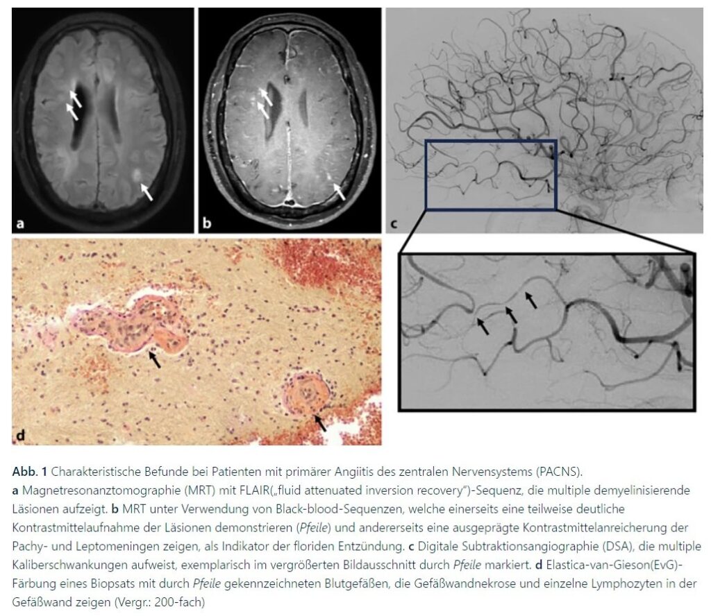
Die serologische Labordiagnostik dient dem Ausschluss von Differenzialdiagnosen, da serologische Parameter bei der auf das ZNS-begrenzten PACNS in der Regel normwertig sind. Die Liquordiagnostik ist obligat zum Ausschluss einer infektiösen Genese und zeigt bei 65–75 % der Patienten mit PACNS eine moderate lymphomonozytäre Pleozytose und/oder eine Eiweißerhöhung. Bei einer Pleozytose > 250/µl sollte hingegen an andere, v. a. infektiöse Erkrankungen gedacht werden. Insbesondere sollte auch mittels PCR („polymerase chain reaction“) und Antikörperindizies aus Liquor und Blut eine Varizella-Zoster-Virus(VZV)-bedingte Vasopathie ausgeschlossen werden. Diese tritt insbesondere bei Kindern häufig auf.
In der Therapie der PACNS wird zwischen Induktions- und Erhaltungstherapie unterschieden. Das Ziel der Induktionstherapie ist die Remission, das Ziel der Erhaltungstherapie der Remissionserhalt und das Verhindern von Rezidiven. Die aktuellen Leitlinien der Deutschen Gesellschaft für Neurologie (DGN) empfehlen eine initiale intravenöse Kortikoidstoßtherapie über 3 bis 5 Tage mit anschließender Oralisierung auf 1 mg/kgKG und nachfolgender schrittweiser Dosisreduktion nach EULAR(European League Against Rheumatism)-Empfehlungen für ANCA-assoziierte Vaskulitiden. Zusätzlich wird eine Induktionstherapie mit Cyclophosphamid für 6 Monate empfohlen. Im Anschluss empfehlen viele ExpertInnen aufgrund des Nebenwirkungsprofils von Cyclophoshamid eine Umstellung auf Azathioprin, Mycophenolat-Mofetil oder Methotrexat. Nach 24-monatiger Rezidivfreiheit unter einer Erhaltungstherapie wird das Erwägen eines Auslassversuchs empfohlen.
Die Riesenzellarteriitis (RZA) ist in Europa die häufigste idiopathische Vaskulitis und tritt ab einem Alter von 50 Jahren mit zunehmender Häufigkeit auf (Peak in der 7. Lebensdekade). Das Lebenszeitrisiko beträgt für Frauen ca. 1 % und für Männer ca. 0,5 %. Als Großgefäßvaskulitis befällt die Riesenzellarteriitis insbesondere die Aorta und ihre direkten Abgänge . Klinisch typisch ist die Manifestation mit neuartigen, akut-subakut einsetzenden, häufig temporal lokalisierten Kopfschmerzen, die meist nicht analgetikaresponsiv sind. Ein pathognomonisches, aber nicht obligates Symptom ist die Kiefer‑/Kau-Claudicatio infolge einer Beteiligung der die Massetermuskulatur versorgenden Äste der Arteria carotis externa . Weitere typische Symptome einer RZA sind Sehstörungen wie Doppelbilder oder (transienter) Visusverlust infolge einer arteriitischen anterioren ischämischen Optikusneuropathie (AION), eine Berührungsempfindlichkeit der Kopfhaut (Kämmschmerzen) und konstitutionelle Allgemeinsymptome wie Gewichtsverlust, Nachtschweiß und Abnahme der körperlichen Leistungsfähigkeit. Schlaganfälle stellen eine klinisch bedeutsame und in ihrer Häufigkeit oft unterschätzte RZA-Manifestation (1,5–7 %) dar. Hierbei ist der Befall des vertebrobasilären Stromgebiets charakteristisch . Insbesondere betroffen ist die A. vertebralis proximal des Abgangs der A. cerebelli posterior inferior (Abschnitte V2, V3, proximale V4) mit einem fließenden Übergang von entzündlich-stenotischem zu physiologischem Gefäßlumen („Slope-Zeichen“)In der klinischen Untersuchung sollte auf eine Druckdolenz, Verhärtung, Schwellung oder eine abgeschwächte Pulsatilität der Arteria temporalis unter Palpation geachtet werden. Laborchemisch sollten das C‑reaktive Protein (CRP) und die Blutsenkungsgeschwindigkeit (BSG oft erhöht mit Werten bis 100 mm in der ersten Stunde, aber auch Werte < 50 mm möglich) bestimmt werden. Normwerte sind gerade bei vorwiegend intrakraniellem Befall möglich und schließen eine RZA nicht aus. Die primär einzusetzende Bildgebungsmethode zur Darstellung entzündlicher Veränderungen der Gefäßwand (oft mit diskontinuierlichem Befall, „skip lesions“) ist die farbkodierte Duplexsonographie der temporalen und axillären Arterien mittels Linearschallköpfen und einer Frequenz von ≥ 15, besser ≥ 18 MHz für die Temporalarterien. Das „Halo-Zeichen“ der Temporalarterie in Form eines zirkulären echoarmen Wandödems gilt als Leitbefund mit einer hohen Spezifität (96 %). Eine Biopsie der Temporalarterie sollte Fällen, in denen die Diagnose trotz hohen klinischen Verdachts nicht allein durch die Bildgebung gesichert werden kann, vorbehalten bleiben. Zu beachten ist, dass die Sensitivität beider Untersuchungen unter Initiierung einer Glukokortikoid(GC)-Therapie innerhalb der ersten Tage stark abnimmt. Dennoch darf die GC-Therapie aufgrund oben geschilderter Komplikationen keinesfalls verzögert werden. Die 18-Fluoro-2-Desoxy-D-Glukose-Positronenemissionstomographie-Computertomographie (18F-FDG-PET-CT) ist geeignet zum Nachweis extrakranieller Gefäßbeteiligungen der RZA und ist in Sensitivität und Spezifität der magnetresonanztomographischen Diagnostik deutlich überlegen. Allgemein konsentierte Diagnosekriterien oder ein trennscharfer Biomarker existieren für die RZA nicht. Die Klassifikationskriterien der American College of Rheumatology (ACR)/European League Against Rheumatism (EULAR) von 2022 wurden für wissenschaftliche Zwecke und nicht für die Diagnosestellung im klinischen Alltag validiert. Die Diagnose wird auf Basis der klinischen Anamnese und Untersuchung, paraklinischer Befunde (Labor und Bildgebung) und je nach Konstellation mittels additiver histopathologischer Untersuchung gestellt. Bestehen typische klinische Befunde sowie ein positiver Bildgebungsbefund (inklusive Duplexsonographie) ist die Diagnose sicher zu stellen . Ist einer der beiden Befunde isoliert positiv, sollte ein weiteres diagnostisches Verfahren in Form weiterer Bildgebungsmodalität oder histopathologischer Untersuchung ergänzt werden. In aktuellen Studien wird an Vorhersagemodellen gearbeitet, die es ermögliche sollen, mittels klinischer Befunde und Ultraschalldiagnostik die RZA zuverlässig von RZA-Mimics trennen zu können.
Entscheidend bei der Therapie der Riesenzellarteriitis ist die Vermeidung jeglicher Zeitverzögerung. Gemäß der ACR-Leitlinie von 2021 besteht die Initialtherapie in der hochdosierten oralen (40–60 mg Prednisolon/Tag) oder im Falle drohender neurologischer (Visus‑)Komplikationen intravenösen (500–1000 mg Methylprednisolon/Tag) GC-Therapie. Da die meisten RZA-PatientInnen ein hohes Risiko für steroidassoziierte Nebenwirkungen aufweisen, sowie aufgrund der meist erforderlichen hohen GC-Dosierungen, sollte eine steroidsparende Therapie in der Regel primär eingesetzt werden. Zugelassen ist hier der Interleukin-6-Rezeptor-Antikörper Tocilizumab (TCZ) in der wöchentlichen subkutanen Gabe von 162 mg. Unter Tocilizumab muss im Falle von Infektionen sowie in der Rezidivdiagnostik beachtet werden, dass es infolge der Inhibition der Akute-Phase-Proteine zu keinem oder nur verzögertem CRP-Anstieg kommt. Ebenfalls wird Methotrexat als steroidsparende Therapie eingesetzt („off-label“). Nach Stabilisierung der Erkrankung sollte das GC gemäß GiACTA-Schema über 52 Wochen (ohne steroidsparende Therapie) oder über 26 Wochen (mit TCZ) ausgeschlichen werden.Abdosierungsschema siehe Tabelle e2 im Onlinezusatzmaterial). Unter GC-Dosierungen > 5 mg/Tag sollten eine Osteoporoseprophylaxe mit täglicher Aufnahme von Vitamin D3 (1000 IE/Tag) und Kalzium (1000–1500 mg/Tag) über die Nahrung oder Supplemente erfolgen. Eine routinemäßige Gabe von Statinen, Thrombozytenfunktionshemmern oder einer oralen Antikoagulation wird nicht empfohlen .Neben der Riesenzellarteriitis können auch die anderen systemischen Vaskulitiden mit einer ZNS-Beteiligung einhergehen. Im Vergleich zur Riesenzellarteriitis haben diese eine wesentlich geringere Inzidenz . Tabelle e1 aus dem Onlinezusatzmaterial gibt eine Zusammenfassung der typischen Symptome, der pathophysiologischen Charakteristika und Therapieempfehlungen.
Infektionen, insbesondere die Sepsis, steigern das Schlaganfallrisiko in einem vergleichsweise kurzen Zeitfenster signifikant. Eine aktuelle Metaanalyse, die Studien mit insgesamt über 120.000 PatientInnen einschließt, zeigt, dass ca. 5 % aller PatientInnen mit Sepsis einen ischämischen Schlaganfall erleiden. Zu den Mechanismen, durch die systemische Infektionen Schlaganfälle verursachen, zählen Embolien durch neu aufgetretenes („symptomatisches“) Vorhofflimmern, hämodynamische Instabilität und das Auftreten infektionsassoziierter Koagulopathien .In einer Analyse von 49.082 PatientInnen mit schwerer Sepsis trat bei 5,9 % neu diagnostiziertes Vorhofflimmern auf. Weiterhin wurde für diese PatientInnen mit sepsisbedingtem, neu aufgetretenem Vorhofflimmern ein fast 4‑fach erhöhtes Schlaganfallrisiko ermittelt . Auch wenn symptomatisches Vorhofflimmern bei kritisch kranken PatientInnen oft vorübergehend ist, kann hierdurch bereits ein erhöhtes Embolierisiko bedingt sein, da sich Vorhofthromben schon innerhalb von 2 Tagen nach Beginn des Vorhofflimmerns bilden können. Die Datenlage zur Antikoagulation bei PatientInnen mit neu diagnostiziertem Vorhofflimmern während einer Sepsis zeigt jedoch ein erhöhtes Blutungsrisiko ohne Risikoreduktion für ischämische Schlaganfälle, sodass im Allgemeinen keine Antikoagulation für PatientInnen mit Sepsis und neu diagnostiziertem Vorhofflimmern empfohlen wird.
Die Inzidenz einer sepsisassoziierten klinischen oder subklinischen Koagulopathie wurde in vorausgehenden Arbeiten auf über 80 % geschätzt. Besonders häufig ist die disseminierte intravaskuläre Koagulation (DIC). Hierbei werden prokoagulante Substanzen wie der Gewebefaktor (TF) freigesetzt, die zur Aktivierung der Blutgerinnung führen und dadurch die Bildung von Thromben im Mikrogefäßsystem oder in größeren Gefäßen verursachen. Die umfangreiche Bildung von Thromben führt wiederum zum Verbrauch von endogenen Gerinnungsfaktoren und Thrombozyten mit der Folge einer Verbrauchskoagulopathie. Endorganschäden resultieren aus reduzierter Perfusion, Thrombosen und/oder Blutungen. Die Diagnose der DIC wird klinisch und laborchemisch gestellt. Die klinische Verdachtsdiagnose ergibt sich bei Patienten, die generalisiertes Sickerbluten aus mehreren intravenösen Katheterstellen oder andere Blutungszeichen aufweisen oder bei Patienten mit unerklärten Thrombosen und einer zugrunde liegenden Erkrankung, die für eine DIC prädestiniert. Die Diagnose einer akuten DIC gilt als gesichert, wenn laborchemische Anzeichen für eine Thrombozytopenie, ein Verbrauch von Gerinnungsfaktoren (z. B. Verlängerung der Prothrombinzeit [PT] oder der partiellen Thromboplastinzeit [aPTT], ein niedriges Fibrinogen) und eine Fibrinolyse (z. B. erhöhter D‑Dimer-Wert) festgestellt werden, vorausgesetzt, es gibt keine andere Erklärung für diese Befunde.
Etwa 20 % der PatientInnen mit bakterieller Meningitis erleiden innerhalb der ersten Wochen nach der Diagnosestellung einen ischämischen Schlaganfall. Neben der bakteriellen Meningitis gehören zu den erregerbedingten Meningitiden auch solche, die durch Viren, Mykobakterien, Pilze oder Protozoen verursacht werden. Infolge der Entzündungsreaktion können bei nahezu allen Formen der Meningitiden sekundär Schlaganfälle auftreten. Die bakterielle Meningitis kann durch das Auftreten typischer klinischer Symptome (Kopfschmerzen, Fieber über 38 °C, Nackensteifigkeit oder Bewusstseinsstörungen) und die Anzahl der Leukozyten im Liquor (über 1000/μl) und/oder den Nachweis bakterieller Mikroorganismen im Liquor diagnostiziert werden. Zerebrovaskuläre Komplikationen, die neben dem ischämischen Schlaganfall auch Hirnvenen- und Sinusthrombosen, Hirnblutungen sowie mykotische Aneurysmen umfassen, tragen zu einem schlechten Outcome der bakteriellen Meningitis bei. Hinsichtlich der zeitlichen Charakteristik kann ein zweigipfeliger Verlauf mit frühen (Tag 3 und 7) und späten (Tag 14) Schlaganfällen beobachtet werden [36
]. Der genaue pathophysiologische Mechanismus zur Entstehung von Schlaganfällen im Kontext der bakteriellen Meningitis ist noch nicht vollständig verstanden, jedoch werden der Nachweis von zerebralem Vasospasmus, Vaskulitis, Gerinnungsstörungen, infektiöser Endokarditis und systemischer Entzündungsreaktionen diskutiert. Inwiefern sich die Entstehungsmechanismen früher und später zerebraler Ischämien unterscheiden, ist zum aktuellen Zeitpunkt ungeklärt. Schlaganfälle infolge einer bakteriellen Meningitis treten häufig disseminiert in beiden Hemisphären lokalisiert auf (Abb. 2a). Interessanterweise konnte in einer Fallserie gezeigt werden, dass die Schlaganfälle vor allem in den Versorgungsgebieten von Arterien auftreten, die sich in der Nähe des Infektionsfokus, wie z. B. einer Mastoiditis, befinden. Das Risiko eines ischämischen Schlaganfalls ist unabhängig vom identifizierten Erreger. Eine Vaskulopathie infolge einer bakteriellen Meningitis lässt sich am sensitivsten anhand multipler intraduraler Gefäßverengungen in der Angiographie nachweisen (Abb. 2b).
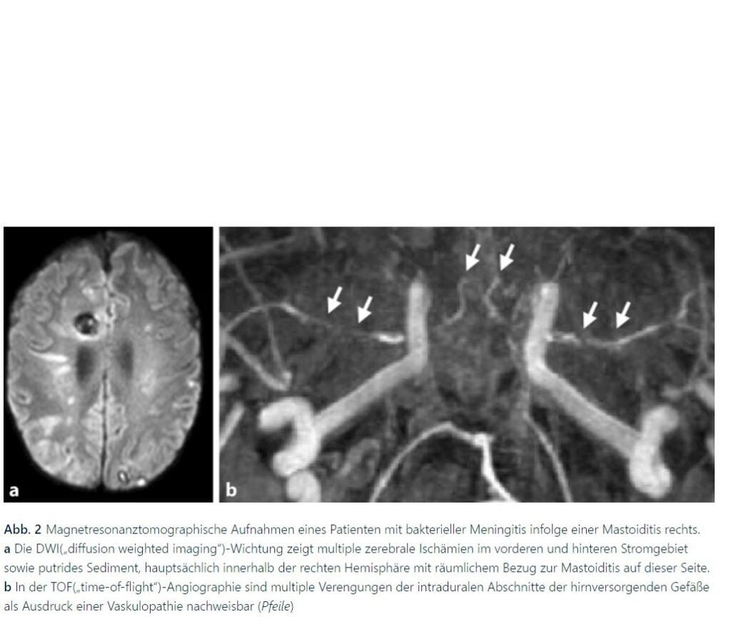
Für den klinischen Alltag relevant ist, dass das Auftreten eines Schlaganfalls mit einer höheren Sterblichkeit im Krankenhaus und einem höheren Grad an persistierenden neurologischen Beeinträchtigungen im Vergleich zu PatientInnen ohne Schlaganfall einhergeht. Eine spezifische Therapie für Schlaganfälle infolge bakterieller Infektionen der Hirnhäute existiert nicht. Es wird primär die bakterielle Infektion basierend auf den aktuellen Leitlinienempfehlungen in Form einer empirischen Antibiotikagabe behandelt. Es gibt bislang keine datengestützten Empfehlungen für die akute Behandlung von Schlaganfällen im Zusammenhang mit einer bakteriellen Meningitis. Prinzipiell ist eine mechanische Thrombektomie möglich, von einer intravenösen Lysetherapie sollte in Anbetracht des möglicherweise erhöhten Blutungsrisikos eher abgesehen werden. Es gibt bisher zwar keine gesicherten Daten für den Einsatz von Acetylsalicylsäure (ASS) zur Sekundärprophylaxe nach vaskulitischem Schlaganfall, jedoch würde sich diese Prophylaxe pragmatisch empfehlen.
Weiterhin hervorzuheben sind zerebrovaskuläre Komplikationen infolge einer Borrelieninfektion. Die Lyme-Borreliose ist eine durch Zecken übertragene multisystemische Infektionskrankheit, die in erster Linie durch drei pathogene Spezies der Spirochäten Borrelia (B. burgdorferi, B. afzelii und B. garinii) verursacht wird. Das Nervensystem ist nach der Haut und den Gelenken das mit 10–15 % am dritthäufigsten betroffene Organsystem. Die Lyme-Neuroborreliose präsentiert sich in der Regel als Symptomtrias aus Meningitis, kranialer Neuritis und Radikuloneuritis, kann aber auch komplizierte Verläufe mit Vaskulitis der Hirngefäße aufweisen. Eine assoziierte Vaskulitis wird in der Literatur mit einer Häufigkeit von 0,3–1 % beschrieben und geht mit einem besonders hohen Schlaganfallrisiko einher. Die leitlinienkonforme Behandlung der Lyme-Neuroborreliose umfasst eine 2‑ bis 3‑wöchige Antibiotikatherapie, in der Regel mit intravenösem Ceftriaxon oder oralem Doxycyclin. Die Deutsche Gesellschaft für Neurologie empfiehlt, bei zerebrovaskulären Manifestationen eine additive Behandlung mit Steroiden und/oder Thrombozytenaggregationshemmern in Betracht zu ziehen. Falls die Krankheitsprogression mittels Antibiotika und Steroiden nicht ausreichend kontrolliert werden kann, sollte eine zusätzliche immunsuppressive Therapie mit Cyclophosphamid evaluiert werden. Hinsichtlich der Akutbehandlung eines Schlaganfalls im Kontext einer Lyme-Neuroborreliose gibt es bisher keine systematischen Arbeiten. In der Literatur sind Einzelfälle zur endovaskulären Behandlung eines vaskulitischen Gefäßverschlusses bei Lyme-Neuroborreliose beschrieben .
Die bakterielle Endokarditis bezeichnet eine bakterielle Infektion der endokardialen Herzoberfläche, vor allem der Aorten- und Mitralklappe. Ihre Inzidenz ist in den letzten 30 Jahren weltweit von etwa 9,9 auf etwa 13,8 pro 100.000 Einwohner angestiegen. Gleichzeitig hat auch die Mortalität zugenommen. Erst 2023 sind die Duke-Kriterien zur verbesserten Diagnosestellung revidiert worden und beinhalten seitdem weitere diagnostische Mittel wie die Herz-CT und die intraoperative Inspektion der Herzklappen. Zur Diagnosestellung bedarf es entweder zweier Hauptkriterien (mikrobiologische Kriterien, bildgebende Kriterien, intraoperative Klappeninspektion) oder eines Hauptkriteriums mit drei Nebenkriterien (vaskuläre Phänomene, immunologische Phänomene, Fieber, Prädisposition, körperliche Untersuchung oder ein mikrobiologischer oder bildgebender Nachweis, der kein Hauptkriterium erfüllt) oder der Erfüllung von fünf Nebenkriterien. Neben direkten Schädigungen des Herzens und Embolien in peripheren Organe (z. B. Niere, Milz) sind ischämische sowie hämorrhagische Schlaganfälle bei bis zu 15–40 % der PatientInnen eine häufige Komplikation der bakteriellen Endokarditis. Ursächlich für die Ischämien sind septische Embolisationen, die sekundär hämorrhagisieren können. Primäre Hämorrhagien werden durch infektiöse (historisch auch mykotische) Aneurysmen und lokale Arteriitiden verursacht. Der Schlaganfall kann dabei das erste Symptom der Endokarditis darstellen. Bereits 4 Monate vor der Diagnosestellung und bis zu 5 Monate nach Endokarditisdiagnose ist das Schlaganfallrisiko relevant erhöht. Gleichzeitig verschlechtert das Auftreten neurologischer Komplikationen das Outcome der Endokarditis Risikofaktoren sowohl für Ischämien als auch für Hämorrhagien sind vor allem Infektionen durch Staphylococcus aureus als auch die Größe der Klappenvegetationen. Bereits ab 10 mm besteht ein relevantes Embolierisiko, bei einer Größe über 30 mm ist, im Vergleich zu kleineren Vegetationen, das Risiko für ischämische Infarkte etwa 2‑fach und für hämorrhagische Schlaganfälle 3,5-fach erhöht. Mitralklappenendokarditiden haben ein höheres Schlaganfallrisiko als solche der Aortenklappe . Beim Auftreten eines ischämischen Schlaganfalles ist die intravenöse Lyse aufgrund des erhöhten Blutungsrisikos kontraindiziert die mechanische Thrombektomie erscheint jedoch sicher und kann gemäß den bekannten Indikationen angewandt werden .Die bakterielle Endokarditis ist eine der wenigen Schlaganfallätiologien, bei denen keine Thrombozytenfunktionshemmung und/oder Antikoagulation indiziert ist. Die Fortführung einer bereits bestehenden Therapie aufgrund anderer Indikationen mit hohem Thrombembolierisiko, wie z. B. Vorhofflimmern oder hochgradiger extra-/intrakranieller Gefäßstenosen, kann jedoch unter Nutzen-Risiko-Abwägung erwogen werden, sofern keine zerebrale Hämorrhagie besteht . Die Leitlinie der European Society of Cardiology aus dem Jahr 2023 empfiehlt zur Prävention weiterer Embolien die operative Versorgung. Hierbei muss das individuelle Embolierisiko gegen das perioperative Risiko abgewogen werden, da intraoperativ eine Antikoagulation erfolgen muss. Bislang liegen hierzu nur Daten aus Beobachtungs-, nicht aber randomisierten Studien vor. Eine frühe operative Versorgung wird empfohlen, solange keine Einblutung besteht und der/die PatientIn nicht bereits präoperativ komatös ist .Auch bei hämorrhagischen Schlaganfällen scheint eine Verzögerung einer indizierten Operation das Outcome zu verschlechtern. Real-world-Daten zeigen, dass eine indizierte operative Versorgung entgegen den Empfehlungen noch zu selten erfolgt.
Schlaganfälle sind eine häufige Komplikation einer bakteriellen Endokarditis. Ursächlich für die Ischämien sind septische Embolien, die sekundär hämorrhagisieren können.
Die primäre Angiitis des zentralen Nervensystems (PACNS) ist eine Vaskulitis unbekannter Ursache. In ihrer Folge treten Schlaganfälle und transiente ischämische Attacken oft mehrzeitig und in unterschiedlichen Gefäßterritorien auf.
Die Riesenzellarteriitis ist eine klinisch bedeutsame und in ihrer Häufigkeit oft unterschätzte Ursache des Schlaganfalls. Klinisch typisch ist die Manifestation mit neuartigen, akut-subakut einsetzenden, häufig temporal lokalisierten Kopfschmerzen, die meist nicht analgetikaresponsiv sind.
Infektionen, insbesondere die Sepsis, steigern das Schlaganfallrisiko signifikant. Zu den ursächlichen Mechanismen zählen Embolien durch neu aufgetretenes Vorhofflimmern, hämodynamische Instabilität und infektionsassoziierte Koagulopathien.
Der Mechanismus zur Entstehung von Schlaganfällen im Kontext der bakteriellen Meningitis ist nicht vollständig verstanden. Diskutiert wird der Nachweis von zerebralem Vasospasmus, Vaskulitis, Gerinnungsstörungen, infektiöser Endokarditis und systemischer Entzündungsreaktionen.
https://link.springer.com/chapter/10.1007/978-3-030-74462-5_7
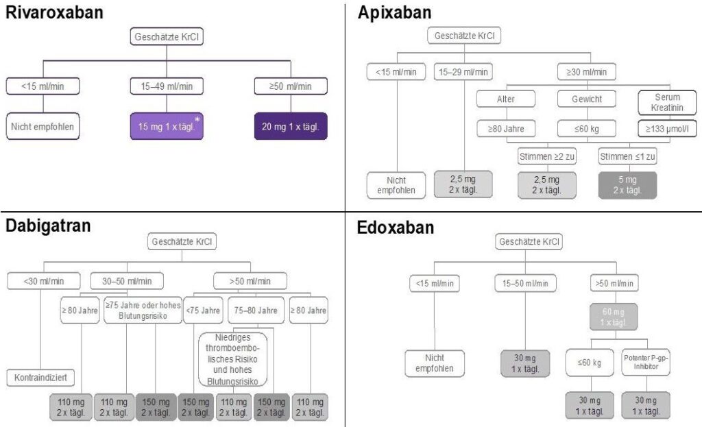
Atrial fibrillation and chronic kidney disease are on the rise worldwide and may coexist in the same patient. The rates of either thromboembolic or hemorrhagic events are increased in patients with kidney disease. Oral anticoagulation therapy, nowadays preferably with direct oral anticoagulants (DOACs), represents the cornerstone for ischemic stroke prevention in high-risk patients. However, all four available DOACs (dabigatran, apixaban, rivaroxaban, edoxaban) are eliminated by the kidneys to some extent. Reduced kidney function facilitates DOACs accumulation and therefore different dose reductions are required, with slight difference between American and European recommendations, particularly in case of severe renal impairment (creatinine clearance <30 mL/min). Overall, the use of DOACs in patients with non-end stage kidney disease and atrial fibrillation is effective similarly to warfarin, showing a better safety profile. Patients with atrial fibrillation on dialysis have been traditionally treated with warfarin, but its effectiveness for stroke prevention in this population is questionable. Some retrospective studies showed promising results with apixaban, but years will be needed for the conclusion of the ongoing randomized clinical trials. In the meantime, the management of stroke risk among patients with atrial fibrillation on dialysis will remain challenging. In patients who underwent renal transplant, DOACs may provide a potentially safer option compared to warfarin. To date, no clinical trial data is available for DOACs use in renal transplant patients, who are treated also with immunosuppressants characterized by a narrow therapeutic index. Co-administration of DOACs with immunosuppressants is a matter of concern given the risk of drug–drug interactions.


An aneurysm is a permanent, localized arterial dilation to more than 50% of the normal diameter. The histopathology of some aortic aneurysms involves medial degeneration (formerly called cystic medial necrosis), characterized by elastic fiber loss from the medial layer, loss of vascular smooth muscle cells, and proteoglycan deposition. Altered microribonucleic acid expression is implicated in increased activity of matrix metalloproteinase (MMP)-2 and -9 , which lyse elastic fibers and break down the extracellular matrix. The media within the aneurysm is also remodeled, with smooth muscle cells oriented randomly, instead of in organized layers parallel to the aortic lumen. Transforming growth factor (TGF)-β, a regulatory cytokine expressed by arterial wall vascular smooth muscle cells, modulates MMP activity. In a mouse aortic aneurysm model, disrupting TGF-β signaling prevented aneurysm formation .There are 2 TGF-β signaling pathways, and both play roles in aneurysm formation . In the canonical pathway, TGF-β binds at the cell surface to its type I or type II receptors. The complex phosphorylates Smad2 or Smad3, resulting in Smad4 recruitment and nuclear translocation, and Smad-dependent gene transcription. In the noncanonical pathway, TGF-β phosphorylates extracellular signal-regulated kinase (ERK), Jun N-terminal kinase, and p38.
Medial degeneration may (at least initially) occur as an adaptive response to wall stress in the aortic dilation zone. Per Laplace’s law, wall tension (T) equals pressure (P) multiplied by radius (R), or T = PR. Aneurysm formation increases wall tension, causing further expansion of the aneurysmal segment. As the aorta enlarges, it loses distensibility. When the tensile limit is reached, dissection may occur, but aneurysms can occur without dissection, and dissections can occur without aneurysms.
Dissection involves disruption of the medial layer, with intramural hemorrhage leading to propagation (especially when the intimal layer is also disrupted) and tracking of blood within the media. Two commonly used classification schemes are used for thoracic aortic dissection: the DeBakey scheme categorizes dissections by the site of origin; and the Stanford scheme categorizes dissections by involvement of the ascending aorta (Central Illustration). In the DeBakey classification, dissections originating in the ascending aorta are subdivided into those that propagate to the aortic arch (type I) and those confined to the ascending aorta (type II), as opposed to those that originate in the descending aorta (type III), regardless of whether they propagate distally or retrograde. The Stanford scheme is divided into type A dissections, which involve the ascending aorta, and type B dissections, which do not, regardless of point of origin .
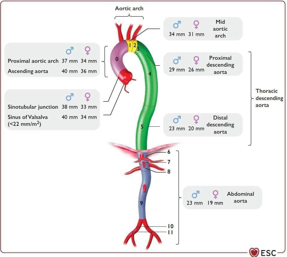
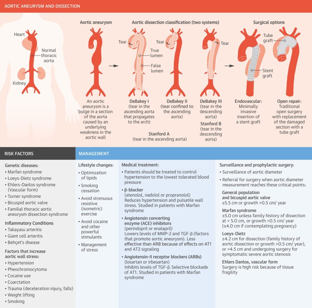
Thoracic Aortic Aneurysm and Dissection.
Judith Z. Goldfinger, Jonathan L. Halperin, Michael L. Marin, Allan S. Stewart, Kim A. Eagle, and Valentin Fuster
JACC. 2014 Oct, 64 (16) 1725–1739