Popeye deformasiyası nədir? Popeye deformasiyası, cizgi filmi qəhrəmanı Popeye-in həddindən artıq böyük bisepsinə bənzəyən şişkin biseps əzələsi ilə xarakterizə olunur. Bu vəziyyət adətən biseps əzələsini çiynə birləşdirən biseps tendonunun uzun başındakı yırtıqdan yaranır. Lakin bəzən bu hal sistem xəstəlikləri zamanıda yarana bilir. 2022 ci ildi Avropa Kardioloji Cəmiyyətinin aylıq dərgisində yayımlanmış məqalədə bir neçə Yapon və İspan alimləri bu haqda çox maraqlı araşdırma aparmışlar. Bu araşdırmaların qısa icmalı original dildə təqdim olunur.
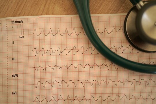
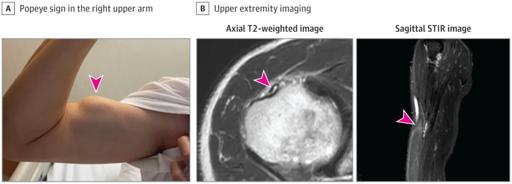
A 76-year-old man with a history of cervical open-door laminoplasty for ossification of the posterior longitudinal ligament presented to our hospital with a symptom of exertional dyspnoea. A 12-lead electrocardiogram revealed I-degree atrioventricular block (PQ interval 262 ms), left axis deviation, and complete right bundle branch block (Panel A). A transthoracic echocardiogram indicated left ventricular hypertrophy with a maximum wall thickness of 17 mm and a left ventricular ejection fraction of 61% (Panel B1). Strain echocardiogram using speckled tracking showed marked diminution of global longitudinal strain with apical sparing (Panel B2). Physical examination found subconjunctival hemorrhage in right eye, several purpuric lesions in upper extremities, and a large bulge on his left upper arm when he flexed his arm, so-called Popeye’s sign, indicating a hallmark of biceps tendon rupture (see Supplementary material online, Video S1; Panel C; arrow). A monoclonal component was not detected by serum and urine immune-electrophoresis and free light chain assays. 99mTc-labelled bone scintigraphy showed grade 3 myocardial uptake (Panel D). Eventually, endomyocardial biopsy and genetic sequencing analysis of the transthyretin gene confirmed the patient as a wild-type transthyretin amyloidosis (Panel E). Popeye’s sign is uncommon, but pivotal to diagnose transthyretin amyloidosis.
Spain Research doünload PDF content A 69-year-old man presented to our cardiology clinic troubled by breathlessness on exertion. He had a history of hypertension, atrial fibrillation, and bilateral carpal tunnel syndrome. Physical examination showed jugular venous distension, bilateral ankle oedema, and a bunching of the right biceps when he flexed his arm—so called Popeye’s sign—indicating rupture of the proximal biceps tendon (figure and video); a rupture was confirmed by an ultrasound scan. Serum N-terminal-pro-B-type natriuretic peptide was 7088 pg/mL (normal range for patients aged 0–74 years is less than 125 pg/mL). A 12-lead electrocardiogram showed low voltage in the limb leads, as well as a pseudo-infarct pattern in the precordial ones (appendix). A transthoracic echocardiogram showed left ventricular hypertrophy with a maximum wall thickness of 20 mm and a left ventricular ejection fraction (LVEF) of 40%. Cardiac MRI showed signs of myocardial infiltration. 99mTechnetium-3,3-diphosphono-1,2-propanodicarboxylic acid (99mTc-DPD) scintigraphy showed grade 3 myocardial uptake (figure). Serum and urine immunoelectrophoresis and free light chain assays ruled out a monoclonal component. Genetic sequencing analysis of the transthyretin gene found no mutations. Tying all the findings together, a diagnosis of heart failure secondary to wild-type transthyretin amyloid (ATTRwt) cardiomyopathy was made. The patient was treated with a combination of diuretics—namely, furosemide 80 mg twice a day, hydrochlorothiazide 12・5 mg once a day, and spironolactone 100 mg once a day—but he did not respond well. He remained symptomatic with advanced functional impairment and persistent signs of systemic venous congestion. ATTRwt, previously known as senile systemic amyloidosis, is a disease that typically affects the heart and tendons of elderly patients. A history of bilateral carpal tunnel syndrome or, less frequently, spontaneous tendon rupture in a patient with heart failure and preserved or mildly depressed LVEF should raise clinical suspicion of the condition. A definitive diagnosis of ATTRwt cardiomyopathy can be confirmed non-invasively with a positive finding on 99mTc-DPD scintigraphy, together with the absence of a monoclonal component in serum and urine samples, and a normal genetic sequencing analysis of the transthyretin gene. Current therapy for patients with ATTRwt cardiomyopathy is limited, with diuretics for relief of cardiac congestion being the mainstay of treatment. However, a recent randomised, placebo controlled trial, published in September 2018, showed a significant reduction in the risk of death and hospitalisation from cardiovascular causes in patients with ATTRwt cardiomyopathy treated with tafamidis, a transthyretin stabilising agent. Tafamidis is expected to be available for clinical use in the near future.
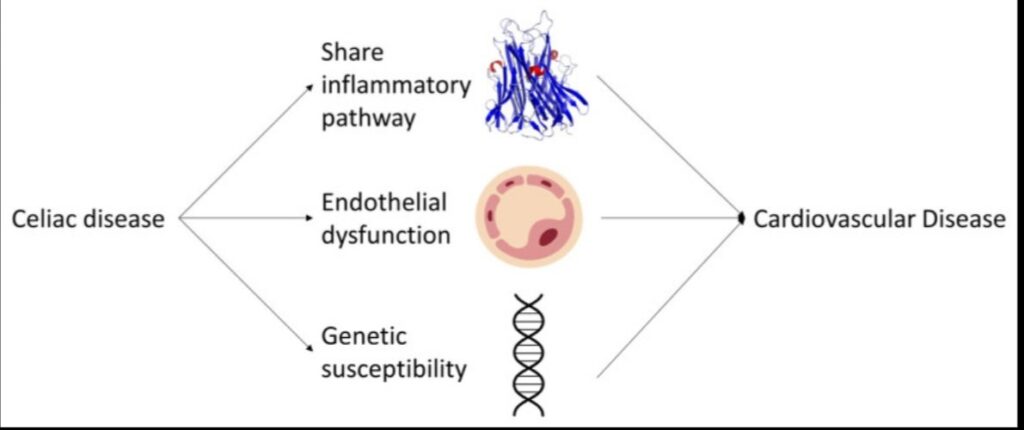
Müasir zamanların xəstəlikləri olan bu iki xəstəlik bir birinə çox oxşasada bəzi fərqləri olmaqdadır. Bu yayınımızda xəstəliklərin ürək damar problemləri ilə əlaqəsini göstərən əlaqə haqqında yazırıq.İngiltərədə təqribən 500000 min insan üzərində aparılmış böyük randomizə olunmuş bir tədqiqat belə əlaqənin olmasını daha aydın göstərir. Seliak xəstəliyinin baş vermə səbəbi autoimmun patologiya nəticəsində yaranmış xroniki iltihab eyni zamanda arteriyalarda aterosklerotik dəyişikliklərin artmasına səbəb olur. Nəticədə kardiovaskulyar xəstəliklərin və gələcəkdə onların ola biləcək ağırlaşmalarının daha sıx üzə çıxması müşahidə olunur. Bu zaman xroniki iltihabdan başqa həmdə genetik əlaqənin də olması prosesin inkişafına təsir edir. Seliak xəstələrinin pəhrizə əməl etmələri isə həm də Kardiovaskulyar xəstəliklərin yaranma riskini azalda bilər.
Association between coeliac disease and cardiovascular disease: prospective analysis of UK Biobank data
Objectives To investigate whether people with coeliac disease are at increased risk of cardiovascular disease, including ischaemic heart disease, myocardial infarction, and stroke.
Design Prospective analysis of a large cohort study.
Setting UK Biobank database.
Participants 469 095 adults, of which 2083 had coeliac disease, aged 40-69 years from England, Scotland, and Wales between 2006 and 2010 without cardiovascular disease at baseline.
Main outcome measure A composite primary outcome was relative risk of cardiovascular disease, ischaemic heart disease, myocardial infarction, and stroke in people with coeliac disease compared with people who do not have coeliac disease, assessed using Cox proportional hazard models.
Results 40 687 incident cardiovascular disease events occurred over a median follow-up of 12.4 years (interquartile range 11.5-13.1), with 218 events among people with coeliac disease. Participants with coeliac disease were more likely to have a lower body mass index and systolic blood pressure, less likely to smoke, and more likely to have an ideal cardiovascular risk score than people who do not have coeliac disease. Despite this, participants with coeliac disease had an incidence rate of 9.0 cardiovascular disease cases per 1000 person years (95% confidence interval 7.9 to 10.3) compared with 7.4 per 1000 person years (7.3 to 7.4) in people with no coeliac disease. Coeliac disease was associated with an increased risk of cardiovascular disease (hazard ratio 1.27 (95% confidence interval 1.11 to 1.45)), which was not influenced by adjusting for lifestyle factors (1.27 (1.11 to 1.45)), but was strengthened by further adjusting for other cardiovascular risk factors (1.44 (1.26 to 1.65)). Similar associations were identified for ischaemic heart disease and myocardial infarction but fewer stroke events were reported and no evidence of an association between coeliac disease and risk of stroke.
Conclusions Individuals with coeliac disease had a lower prevalence of traditional cardiovascular risk factors but had a higher risk of developing cardiovascular disease than did people with no coeliac disease. Cardiovascular risk scores used in clinical practice might therefore not adequately capture the excess risk of cardiovascular disease in people with coeliac disease, and clinicians should be aware of the need to optimise cardiovascular health in this population.Back to top
What is already known on this topic
Evidence is conflicting on whether coeliac disease is associated with a higher risk of cardiovascular disease, with previous research being conducted in small cohorts or using registry data sources with limited sociodemographic and lifestyle data
Previous studies have tended not to take traditional cardiovascular risk factors, such as blood pressure or serum total cholesterol, into account when examining the association between coeliac disease and cardiovascular disease, despite research showing a healthier cardiovascular profile in people with coeliac disease
What this study adds
Individuals with coeliac disease had a lower prevalence of traditional cardiovascular risk factors, such as systolic blood pressure, total cholesterol, and body mass index, but a higher risk of developing cardiovascular disease, than people with no coeliac disease
How this study might affect research, practice, or policy
Cardiovascular risk scores need to adequately account for the elevated risk among people with coeliac disease; people with coeliac disease and their clinicians need to be aware of their higher cardiovascular risk and to take relevant action; and further research is needed to understand the mechanisms underlying these associations
Introduction
Coeliac disease is an autoimmune disorder resulting in an immune reaction to gluten, a protein found in barley, wheat, and rye. This disease occurs in about 1% of the UK population,1 2 although prevalence is increasing, partly due to improved diagnostics.2 Coeliac disease is more common in women and is typically diagnosed in childhood and adolescence or at ages 40-60 years, but can occur throughout the life course.3 Symptoms include diarrhoea, weight loss, anaemia, chronic fatigue, and other intestinal and extraintestinal symptoms.3 Comorbidities are common in people with coeliac disease, with an increased risk of osteoporosis4 and some cancers, including non-Hodgkin’s lymphoma and small intestinal adenocarcinoma,5 as well as anaemias and other autoimmune conditions such as dermatitis herpetiformis.3 4 Treatment for coeliac disease involves following a strict gluten-free diet,3 which helps to alleviate symptoms and reduces the risk of comorbidities but does not eliminate the condition.3
Some studies have reported that coeliac disease is associated with an increased risk of cardiovascular disease, including ischaemic heart disease and stroke.6–14 However, evidence is conflicting15–18 and is largely based on case-control, cross sectional, or longitudinal studies that do not have complete or detailed information on cardiovascular risk factors and other potentially confounding factors. Based on this conflicting evidence, cardiovascular disease is not currently considered a complication of coeliac disease by National Institute for Health and Care Excellence, the body that sets guidelines for clinical practice in the UK.19
Previous studies have tended not to explore the role of traditional cardiovascular disease factors, such as blood pressure or serum total cholesterol, when examining the association between coeliac disease and cardiovascular disease, despite research showing a healthier cardiovascular profile in people with coeliac disease.7 We report the findings on the association between coeliac disease and cardiovascular disease using the UK Biobank data,20 a large scale, prospective cohort study, and assess whether any association is independent of traditional cardiovascular disease risk factors.Back to top
Methods
Study design and participants
UK Biobank is a population based cohort study that recruited about 500 000 adults aged 40-69 years from England, Scotland, and Wales between 2006 and 2010.20 Participants attended a baseline assessment centre where they provided sociodemographic, lifestyle, and detailed self-reported health information via a touchscreen questionnaire and verbal interview, had physical measurements taken, and provided non-fasting blood samples. A series of serum biomarkers relevant to cardiovascular disease (total cholesterol, triglycerides, glucose, low density lipoprotein, HbA1c, and C reactive protein) were measured in all participants using a Beckman Coulter AU5800 clinical chemistry analyser21 and Bio-Rad Variant II Turbo analyser.22
All participants provided consent for ongoing linkage to electronic health records to allow longitudinal collection of health events. Hospital inpatient records were obtained from Hospital Episode Statistics for England (available from 1996 until 30 September 2021), Scottish Morbidity Record for Scotland (available from 1981 until 31 July 2021), and Patient Episode Database for Wales (available from 1998 until 28 February 2018). Death records were obtained from NHS Digital (England and Wales; available from the start of recruitment in 2006 until 30 September 2021) and NHS Central Register, National Register of Scotland (Scotland; available from the start of recruitment in 2006 until 31 October 2021).
Assessment of coeliac disease
Coeliac disease status at recruitment was ascertained by use of a combination of self-reported and hospital inpatient diagnosis. For self-report, participants were asked during the touchscreen questionnaire at baseline: “Has a doctor ever told you that you have had any serious medical conditions or disabilities?” If they answered “yes,” then this answer was followed up by a trained nurse during a verbal interview, with coeliac disease one of the medical conditions reported. For the hospital inpatient records, coeliac disease diagnosis was based on International Classification of Diseases (online supplemental table 1) prior to the baseline assessment.
Assessment of cardiovascular disease
Incident cardiovascular disease was ascertained from the hospital inpatient and death records. Secondary outcomes were ischaemic heart disease, myocardial infarction, and stroke. A composite cardiovascular disease outcome (ischaemic heart disease, myocardial infarction and stroke combined) was derived as the main outcome (codes available in online supplemental table 1). Any participant self-reporting heart disease or stroke at baseline, or who had cardiovascular disease diagnostic codes in the hospital inpatient data prior to recruitment, was excluded from the analysis (codes available in online supplemental table 1).
Covariates and cardiovascular risk factors
Socioeconomic status was measured using the Townsend deprivation index score, assigned to each participant using their home postcode at recruitment (grouped into five groups (quintiles)).23 The assessment centre location was used to derive region (England (North East, North West, Yorkshire and Humber, East Midlands, West Midlands, South, South West, London), Scotland, Wales). Ethnicity (white, non-white), highest education level (GCSE, A Levels, higher education, none of the above), alcohol consumption (never, special occasions only, one to three times per month, one to two times per week, three to four times per week, daily), smoking status (never, previous, current), family history of heart disease (none, one parent, both parents), medication use (antihypertensive and cholesterol lowering; yes or no) and physical activity were self-reported via the touchscreen questionnaire. The International Physical Activity Questionnaire24 guidelines were used to derive low, moderate, and high self-reported physical activity levels. Body mass index was derived from weight (measured by the Tanita BC4 18MA body composition analyser) and standing height measured at recruitment. Systolic and diastolic blood pressure was measured twice, with at least one minute between measurements, using an Omron 705 IT electronic blood pressure monitor with the participant in a seated position. For the prospective analysis, a mean of the two readings was derived and categorised into quintiles. Diabetes was self-reported at the baseline assessment visit or a hospital diagnosis prior to recruitment (codes available in online supplemental table 1). Furthermore, diabetes was categorised into type 1, 2, and unspecified, because the association between coeliac disease and diabetes is type specific (see online supplemental methods and supplemental table 1). For the prospective analysis, the composite definition was used due to small numbers of participants with type 1 diabetes. For the prospective analysis, total cholesterol, blood glucose, systolic blood pressure, triglyceride, low density lipoprotein, C reactive protein, and HbA1c were grouped into fifths. For all variables where data was missing, an additional category for missing data was included. A cardiovascular risk score was generated based on a modified version of the American Heart Association’s Life’s Simple Seven risk score (LS7).25 26 This score was selected because it is well validated,26 27 the data required to construct it is readily available in UK Biobank, and has previously been used in UK Biobank research25 The score used in these analyses comprise six known cardiovascular risk factors (smoking, physical activity, total cholesterol, diabetes status, blood pressure, body mass index; diet was omitted as the data required to derive fibre and sodium intake were not available) to characterise a person’s cardiovascular risk profile as ideal, intermediate or poor (see online supplemental methods and supplemental table 2 for the scoring method).
Statistical analysis
Participants with prevalent cardiovascular disease (n=33 364) were excluded from the analysis. Of the serum biomarkers of interest (total cholesterol, glucose, HbA1c, triglyceride, low density lipoprotein, and C reactive protein), the distribution of C reactive protein and triglycerides were right skewed and the data were log transformed to reduce departure from normality. Demographic and cardiovascular disease risk factor variables included in the LS7 were cross tabulated by coeliac disease status. Means and proportions were adjusted to the baseline distribution for age, sex, Townsend score, education, and ethnicity to allow better comparison of co-variates between participants with and without coeliac disease.28 All variables of interest were examined for missing data. The number of variables with missing data was calculated for each participant, and this number was examined to investigate the proportion of participants missing data on more than two variables. Most (12 of 16) of the variables had some missing data, with this ranging from 0.1% of participants (Townsend Score) to 14.5% (non-fasting glucose) (online supplemental table 3). Fewer than 3.5% of participants had missing data for more than two variables (online supplemental table 4).
Incident rates for cardiovascular disease were calculated and stratified by risk score category (ideal, intermediate, and poor). Cox proportional hazards models were used to calculate the hazard ratio for the cardiovascular disease outcomes of interest. Three models were built: model A was adjusted for standard covariates (adjusted for sex, Townsend score, education, region, year of birth, year of recruitment, and ethnicity); model B was further adjusted for lifestyle factors (as model A plus smoking, alcohol consumption, and physical activity); and model C was further adjusted for cardiovascular risk factors (as model B plus family history of heart disease, total cholesterol, glucose, antihypertensive use, cholesterol lowering medication use, and diabetes). The censoring date was the date of first diagnosis of an outcome of interest (either cardiovascular disease, ischaemic heart disease, stroke, or myocardial infarction), date of death, date of lost to follow-up, or last date of available hospital record, whichever came first. The proportional hazards assumption was examined graphically via log-log plots. The models were stratified for year of birth, year of recruitment, alcohol consumption, and body mass index because these factors violated the proportional hazards assumption. Age was used as the underlying time variable. Where models included covariates with missing data, a separate category of the given covariate was used; multiple imputation of missing data by chained equation, with 35 iterations,29 was performed as sensitivity analysis (model D).
We conducted secondary analyses to investigate whether increased inflammation mediated the association between coeliac disease and cardiovascular disease risk by including C reactive protein as a further covariate in the model. Total cholesterol and glucose concentrations were included in the main models to replicate the inclusion in the LS7 risk score. To investigate if the association remained the same with other known cardiovascular biomarkers, total cholesterol and glucose concentrations were replaced with triglyceride, low density lipoprotein, and HbA1c as variables. Participants provided a date of first diagnosis, which was converted to age at diagnosis by UK Biobank. Using this date and the date of recruitment, time since coeliac disease diagnosis was categorised as no coeliac disease, coeliac disease for <10 years, and coeliac disease for ≥10 years at baseline. Models A, B, and C were repeated with time since coeliac disease diagnosis as the exposure and cardiovascular disease as the outcome.
To further investigate the differences in incidence rates of cardiovascular disease by LS7, a joint effects analysis and analyses within groups were undertaken. A joint effects variable was generated that categorised participants by both their coeliac disease status and LS7 category, (no coeliac disease/ideal, no coeliac disease/intermediate, no coeliac disease/poor, coeliac disease/ideal, coeliac disease/intermediate, coeliac disease/poor). Cox proportional hazards modelling was used to investigate the joint effects of coeliac disease status and LS7, adjusted as for model A (as the risk score takes the other potential confounders and effect modifiers into account). For a within group analysis, model A was stratified by LS7 risk score category.
We used Stata SE version 17 (StatCorp, College Station, TX) for our analyses.
Patient and public involvement
Participants were not involved in the development of the specific research question or outcome measures for this article. Participants were involved in developing the ethics and governance framework for UK Biobank and have been engaged in the progress of UK Biobank through follow-up questionnaires and additional assessment visits. UK Biobank keeps participants informed of all research output through the study website (https://www.ukbiobank.ac.uk/explore-your-participation), participant events, and newsletters.Back to top
Results
Of 502 459 UK Biobank participants, 469 095 were included in this study (33 364 participants had prevalent cardiovascular disease and were excluded from further analyses) and 2083 participants had coeliac disease. During follow-up, 1236 (0.3%) of participants were lost to follow-up (owing to leaving the UK), 1435 (0.3%) were diagnosed with coeliac disease, and 24 707 (5.3%) participants died.
Compared with people who do not have coeliac disease, participants with coeliac disease were more likely to be women (55.8% v 71.5%) and of a white ethnic background (94.6% v 98.4%) (table 1). Coeliac disease participants also had a lower body mass index, consumed less alcohol, less likely to smoke, more likely to report a family history of heart disease, had lower total cholesterol and C reactive protein concentration, lower mean systolic blood pressure, were less likely to be diagnosed with type 2 diabetes, more likely to be diagnosed with type 1 diabetes and less likely to use cholesterol lowering or antihypertensive medication, when adjusted for age, sex, socioeconomic status, education, and ethnicity (table 2). Compared with those without coeliac disease, those with coeliac disease were more likely to have a so-called ideal cardiovascular risk score (23.3% v 14.3%), and were less likely to have a poor risk score (5.0% v 8.6%) (table 2). The unadjusted distribution of baseline characteristics were similar to the adjusted results (online supplemental table 5).
Table 1View inline
•Open as popup
Baseline characteristics of the 469 095 participants included in the main analysis, by pre-existing coeliac disease. Data are number (%), unless otherwise specified
Table 2View inline
•Open as popup
Distribution of lifestyle and cardiovascular risk factors, by pre-existing coeliac disease
Over a median follow-up of 12.4 years (interquartile range 11.5-13.1), 40 687 cardiovascular disease events, 33 556 ischaemic heart disease events, 8859 stroke events, and 12 853 myocardial infarction events occurred.
Participants with coeliac disease had a higher absolute incidence of cardiovascular disease compared with people with no coeliac disease (9.03 per 1000 person years (95% confidence interval 7.90 to 10.31) v 7.37 (7.30 to 7.44), P for incidence rate difference <0.001).
Coeliac disease was associated with a 27% increased risk of cardiovascular disease compared with participants who did not have coeliac disease (hazard ratio 1.27 (95% confidence interval 1.11 to 1.45)). The results remained similar after further adjustment for lifestyle factors (1.27 (1.11 to 1.45)), whereas adjustment for cardiovascular risk factors increased the strength of the association (1.44 (1.26 to 1.65)) (table 3). The pattern of associations were similar for ischaemic heart disease and myocardial infarction (table 3). Fewer events of stroke occurred than for other endpoints, and no association was noted between coeliac disease and the risk of stroke.
Table 3View inline
•Open as popup
Association between coeliac disease and cardiovascular disease (CVD) outcomes, with progressive adjustments
The findings remained similar after using multiple imputation to account for missing data (model D online supplemental table 6). In a secondary analysis that aimed to investigate whether increased inflammation could explain the increased risk of cardiovascular disease, ischaemic heart disease, or myocardial infarction, all models were further adjusted for C reactive protein. No attenuation of the results was identified (model C plus log C reactive protein: hazard ratio for cardiovascular disease 1.39 (95% confidence interval 1.21 to 1.60), P<0.001; ischaemic heart disease 1.49 (1.28 to 1.74), P<0.001; and myocardial infarction 1.60 (1.25 to 2.05), P<0.001). No difference was noted in the results when triglyceride, low density lipoprotein, and HbA1c concentrations were included rather than total cholesterol and glucose concentrations (cardiovascular disease 1.39 (1.21 to 1.60), P<0.001; ischaemic heart disease 1.51 (1.30 to 1.76), P<0.001; myocardial infarction 1.66 (1.31 to 2.10), P<0.001; and stroke 1.09 (0.80 to 1.50), P=0.58).
Evidence of a dose-response association between time since coeliac disease diagnosis and risk of cardiovascular disease was reported. Compared with people who do not have coeliac disease, people who had coeliac disease for less than 10 years had a 30% increased risk (model C 1.30 (95% confidence interval 1.07 to 1.57)), and people who had coeliac disease for 10 years or more had a 34% increased risk (1.34 (1.11 to 1.61), P value for trend<0.001; online supplemental table 7).
When investigating the potential joint effects of coeliac disease and cardiovascular risk score and incident cardiovascular disease, people with coeliac disease and an ideal risk score had more than 60% increased risk (hazard ratio 1.64 (95% confidence interval 1.14 to 2.35) P=0.007; figure 1) compared with people with no coeliac disease and an ideal risk score of cardiovascular disease. Participants with an intermediate risk score and coeliac disease had 2.3 times an increased risk (2.30 (1.93 to 2.74), P<0.001) and people with a poor risk score and coeliac disease had almost three times an increased risk (2.89 (1.71 to 4.88), P<0.001), compared with those with an ideal score and no coeliac disease. The increased risk noted with a poor risk score compared with ideal risk was similar in people with coeliac disease (2.89 (1.17 to 4.88)) and in people who do not have coeliac disease (2.78 (2.65 to 2.92)), indicating that coeliac disease did not further amplify the risk of cardiovascular disease in people who are already at high risk of cardiovascular disease (figure 1), but the number of participants in this group was small (table 4). As such, within the ideal and intermediate risk score categories, the hazard ratios for incident cardiovascular disease were 1.57 ((95% confidence interval 1.10 to 2.35), P=0.01) for those with coeliac disease compared with 1.35 ((1.14 to 1.60), P<0.001) for those who do not have coeliac disease (table 4); however, no association was noted for for people in the poor risk score category (1.03 (0.61 to 1.74)).
Risk of incident cardiovascular disease by coeliac disease and cardiovascular risk score. Hazard ratios adjusted for region, sex, Townsend score, education, year of birth, year of recruitment, and ethnicity, with age as underlying time variable. The vertical line represents hazard ratio of 1. Risk score was defined using the American Heart Association’s Life’s Simple Seven score. CI=confidence interval
Table 4View inline
•Open as popup
Analysis within groups for association between coeliac disease and cardiovascular disease, within risk score categoryBack to top
Discussion
Principal findings
To our knowledge, this study is the largest longitudinal study to investigate the association between coeliac disease and cardiovascular disease that allowed for robust adjustment of potential confounders. We adjusted for including a wide range of lifestyle, medical, and cardiovascular risk factors (including biomarkers), showing that the increased risk is not explained by traditional cardiovascular risk factors. Furthermore, we were able to investigate dose-response by time since coeliac disease diagnosis. We found the risk of cardiovascular disease increased as time since coeliac disease increased, suggesting that coeliac disease might increase the risk of cardiovascular disease, with the longer the exposure, the higher the risk. Use of self-reported health data and hospital inpatient data from the mid-1990s meant most cases of coeliac disease are likely to have been captured over time.
Comparison with other studies
Our findings complement previous research that reported that specific cardiovascular disease conditions were over-represented in individuals with coeliac disease,11 13 14 including a UK Biobank study that investigated across all diseases in a hypothesis-free approach.12 We build on these findings by accounting for the role of cardiovascular health in the analysis. Previous research investigating coeliac disease with risk of cardiovascular disease has produced conflicting findings, showing either an increased risk6–14 or no association.15–18 However, studies vary in adjustment, with some studies having minimal adjustments (eg, for age, sex, and socioeconomic status)6 12 15 16 and others further adjusting for comorbidities (such as other autoimmune diseases and hypertension).8 10 11 13 18 Two meta-analyses have been undertaken, one identifying an increased risk of incident stroke,9 and the other identifying an increased risk of death from stroke.17 Neither identified an association between coeliac disease and incident cardiovascular disease, myocardial infarction, or cardiovascular disease death.11 18 In both meta-analyses, the included studies were of low quality, with little adjustment for lifestyle and health related confounders. The authors of both meta-analyses note that large prospective studies are needed, which adequately adjust for confounding, to investigate the association between coeliac disease and cardiovascular disease. The absence of association with stroke and coeliac disease identified in this study could be due to a lack of power because the number of stroke events in the coeliac disease participants was low. To our knowledge, no previous study has taken into account cardiovascular risk score or biochemical risk factors for cardiovascular disease, such as cholesterol, although patients with coeliac disease have a better cardiovascular risk profile than do people who do not have coeliac disease.7 Our research has highlighted that the increased risk of cardiovascular disease is more marked in those with an ideal cardiovascular risk profile.
Numerous autoimmune diseases have been found to be associated with an increased risk of cardiovascular disease.14 One hypothesis for the increased risk is that increased systemic inflammation associated with coeliac disease could subsequently increase the risk of cardiovascular disease.30–32 In our study, the results remained similar when adjusting for C reactive protein, a marker of systemic inflammation. C reactive protein might be a poor marker for inflammation in patients with coeliac disease,7 33 as found in other auto-immune diseases,34 although small case control studies have identified an increased C reactive protein in patients with coeliac disease.32 35 Systemic inflammation is known to trigger atherosclerosis,36 and previous studies have found that patients with coeliac disease have increased intima-medial thickness, reduced elasticity of the ascending aorta, and endothelial dysfunction.35 37 38 A similar association with increased risk of atherosclerosis has been identified in other auto-immune disorders, such as rheumatoid arthritis and systemic lupus erythematosus.39 40 Some studies have shown that a gluten-free diet reduces inflammation and cardiovascular disease in patients with coeliac disease.6 41 Our study cannot investigate whether adherence to a gluten-free diet reduced inflammation and, therefore, whether this factor affected the risk of cardiovascular disease in patients with coeliac disease.
A further hypothesis is that the consumption of a gluten-free diet might increase the risk of cardiovascular disease because gluten-free foods are higher in saturated fats, sugar, and salt,42 or because a gluten-free diet limits consumption of complex whole grains.43 Previous studies have identified changes in cardiovascular biomarkers (such as body mass index, total cholesterol, and triglycerides) after implementing a gluten-free diet, but these changes did not indicate to a better or worse cardiovascular profile.44 We were unable to explore the direct affect of diet (due to only having data for a wheat-free diet, and the small proportion of participants with coeliac disease reporting not eating a wheat-free diet). However, any downstream effect of diet is unlikely to be reflected in the cardiovascular risk factors we investigated in the current study; a Cochrane review found no association between gluten-free diet and cardiovascular disease risk.45 Another potential mechanism for the increased risk noted is through micronutrient deficiencies. Micronutrient deficiencies (such as vitamins A, B, D, and E) have been associated with cardiovascular disease outcomes, although evidence is conflicting.46 Micronutrient deficiencies can occur in patients with coeliac disease who do not adhere to a gluten-free diet (due to malabsorption from villi atrophy), but also occurs in people who do adhere to a gluten-free diet, due to the inadequate micronutrients in a gluten-free diet.47 However, micronutrient deficiencies are common in the UK population (especially among women),48 49 and so is unlikely to fully explain the increased risk of cardiovascular disease seen in coeliac disease participants identified in this study.
Strengths and limitations
The study was limited in that the traditional cardiovascular disease risk factors (including biomarkers and blood pressure) were only measured in the full sample at recruitment, and hence, we could not investigate the impact of changes in cardiovascular disease risk factors over time on risk. Coeliac disease status was ascertained by use of a combination of self-reported and hospital inpatient data. This process might have led to an under ascertainment of cases due to participants not self-reporting a diagnosis and not having a diagnosis recorded in their hospital record (because of how diseases are recorded in Hospital Episode Statistics). As a result of the amount of missingness for some variables (ie, physical activity and some biomarker data), missing data were maintained in the models to ensure adequate power. Although the inclusion of missing data has the potential to bias the results, these variables were covariates and fewer than 3.5% of participants had missing data for more than two variables. Furthermore, multiple imputation showed no change in results when accounting for the missing data, suggesting no bias was introduced by use of this method. A gluten-free diet was not taken into account as the diet questionnaire only asked whether a wheat-free diet was followed (gluten is identified in other grains, such as barley and rye) and the number of participants with coeliac disease who reported not following a wheat-free diet was small, so the impact of a gluten-free diet could not be assessed. The association could be due to ascertainment bias because people with a chronic disease, such a coeliac disease, are more likely to have a hospital record, and, as such, are also more likely to have a cardiovascular disease recorded in their hospital record.50 51 However, analyses that were restricted to myocardial infarction as the outcome (which is predominantly diagnosed in hospital) were very similar to those for cardiovascular disease as a whole, suggesting that ascertainment bias is unlikely to have influenced the results in any meaningful way.
Although this study is large, it lacked power to assess specific cardiovascular disease subtypes and so joint effects of cardiovascular risk score and coeliac disease status on cardiovascular disease subtypes was not possible. Possible conclusions were also not made for the lack of difference in risk for people with a poor cardiovascular risk score (which could reflect a lack of power in this subgroup or could indicate that the cardio-protective therapies that are likely implemented for those with a poor risk score are protective against cardiovascular disease in those with coeliac disease). As with all observational studies, residual confounding likely remains and causality cannot be determined. As UK Biobank is a volunteer based cohort, the so-called healthy volunteer effect might affect consequent findings,52 which could explain the lower prevalence of coeliac disease noted in this study compared with the general population. Additionally, the absolute incidence rates reported in this paper are unlikely to be generalisable to the wider population.53 Nevertheless, the internal associations identified are likely to be generalisable to the population as a whole owing to the wide heterogeneity of risk factors studied.53
Conclusion
This study highlights the importance of cardiovascular disease as a potential complication of coeliac disease. Further research into the drivers and mechanistic pathways of this association is warranted. In addition, an investigation is warranted into the extent to which any risk reduction is reported by adherence to a gluten-free diet in people with coeliac disease, or whether a gluten-free diet itself contributes to the increased risk identified. Furthermore, consideration should be given to inclusion of coeliac disease as a risk factor in cardiovascular disease risk prediction models, such as the QRISK model,54 which currently includes other autoimmune conditions (systemic lupus erythematosus and rheumatoid arthritis) as risk factors. Given the increased rates of cardiovascular disease reported in people with coeliac disease who have an ideal and moderate cardiovascular disease risk score, clinicians should make patients with coeliac disease aware of their elevated risk, and work with their patients to optimise their cardiovascular health.
People reporting cold drinks or foods as triggers for their atrial fibrillation (Afib) were invited to a survey.
Approximately half the cohort reported reducing their Afib episodes by avoiding cold ingestion, with methods including waiting for drinks to warm to room temperature and eliminating straw use.
Healthcare providers often held a dismissive attitude towards the so-called “cold drink heart” phenomenon, the study showed.
Avoiding ice water, smoothies, and ice cream seemed to work as a lifestyle change for some with atrial fibrillation (Afib or AF), according to the first cross-sectional survey of people with “cold drink heart” (CDH).
Among people who claimed to have ever had symptomatic Afib triggered by cold ingestion, 51.5% reported that their Afib episodes occurred only following cold drink or food consumption (as opposed to other triggers). In these patients, avoidance of cold ingestion reduced or eliminated their Afib episodes with 100% effectiveness (as opposed to 72.4% in people who also had a history of non-CDH Afib).
“When asked about avoidance, respondents reported several effective behavioral modifications in lieu of complete avoidance, such as reducing speed of ingestion or avoiding rapid gulping, eliminating straw use, allowing drinks to warm to room temperature, or warming liquids in their mouth before swallowing,” reported David Vinson, MD, of Kaiser Permanente (KP) Northern California in Pleasanton, and colleagues in the Journal of Cardiovascular Electrophysiologyopens in a new tab or window.
With their survey, the investigators had taken the first step to study the cold drink-Afib link more systematically, beyond the initial case reports.
The survey included 101 people who self-reported cold ingestion-triggered symptomatic Afib or atrial flutter at KP Northern California emergency departments (n=39) and an additional cohort of non-KP patients who had contacted the research team, unsolicited, offering their experience with CDH (n=62).
“We did this study because for decades there have been people telling their healthcare providers that cold foods and drinks trigger their atrial fibrillation episodes — but many providers have dismissed this possibility,” said Vinson in a press release. “Yet, the more patients with atrial fibrillation are asked about — or read about — this trigger, the more often we hear, ‘Yes, that’s happening to me, too.’”
“Although the majority of the people we surveyed said their atrial fibrillation was associated with cold ingestion, it was actually rare that eating or drinking something cold always precipitated an atrial fibrillation episode,” Vinson noted. “In other words, most people with cold drink heart were often able to eat cold food or drinks without developing symptoms of atrial fibrillation. This shows how unpredictable the condition can be and why it’s been hard for some patients to identify these triggers.”
He and his study co-authors urged greater clinician awareness of CDH in the setting of Afib, citing one estimate that 5-10% of people with paroxysmal Afib may have cold drinks or foods as a trigger.
In their present report, patients said they had gotten a range of reactions from professionals when they shared their experiences: 52.4% of respondents reported dismissive attitudes from one or more healthcare providers, while some actually said they learned about the phenomenon from a physician.
In practice and in research, alcohol is more established as a dietary trigger of Afib.
“Alcohol ingestion has been recently identified as a trigger of discrete AF events, but only after a delay of 3-12 [hours]. This hours‐long delay contrasts with the relative immediacy of cold drink triggers, which precipitate AF within seconds to minutes,” wrote Vinson’s group.
Vinson and colleagues recruited a study cohort that was 75% men with a median age of 56 years, a median CHA₂DS₂‐VASc score of 1, and 25.7% on anticoagulants. Patients said they had been 44.5 years old at CDH onset.
When asked which rhythm was triggered by cold ingestion, 74.3% of patients reported only Afib, 15.8% said both Afib and flutter, 3.0% flutter only, while 7.0% said they were unsure.
The KP cohort underwent chart review for ECG confirmation of their Afib. As for the outside cohort, a documented diagnosis of AF or atrial flutter was required but these patients did not have to share their medical records.
Study authors noted that 36.5% of those surveyed reported that Afib was often triggered by cold ingestion soon after physical activity, and some reported that this was always the case for their CDH episodes.
“While the underlying mechanism could not be elucidated by the current study, an exacerbation after exercise suggests a vagotonic effect. Indeed, heightened vagal tone appears to trigger AF, and esophageal stimulation such as with a cold drink, may acutely increase such an autonomic response,” Vinson’s group surmised.
“Alternatively, as the esophagus lies directly behind and often in contact with the posterior left atrium, direct cold mechanical irritation of the left atrium may also be responsible, which also could be exacerbated post-exercise due to the relative increase in vagal tone,” the investigators continued.
Rapid gulping or swallowing was reportedly also more likely to provoke Afib, the survey found.
Vinson and colleagues acknowledged that the study was not designed to prove causality and that the survey responses may have been inaccurate. There was likely some degree of selection bias affecting the study, as well.
“Further study will be required to assess the prevalence of CDH among an unselected population of AF patients, and importantly, the generalizability of our findings around the effectiveness of cold drink avoidance and other behavioral modifications in reducing AF recurrence,” they wrote.
People living with type 2 diabetes (T2D) who also have albuminuria are up to four timesopens in a new tab or window more likely to die from a cardiovascular (CV) event, five times more likely to be hospitalized for heart failure, and three times more likely to have a heart attack compared to people with T2D alone. In fact, a 2025 prospective analysis in Austrian patients stated that albuminuria may be on paropens in a new tab or window with the same risk as a prior heart attack when it comes to predicting a future heart attack.
Even though albuminuria can be detected through a common and inexpensive urine test — urine albumin-to-creatinine ratio (UACR) — we aren’t screening for it as often as we should or treating it adequately.
If UACR is ≥30 mg/g for at least three months, the patient is considered to have albuminuriaopens in a new tab or window, which is not only important for identifying CV risk but also an early sign of chronic kidney disease (CKD). Once a patient is diagnosed with albuminuria, UACR testing frequency should increase.
In reality, only about halfopens in a new tab or window of T2D patients have their UACR checked once a year. What’s more, those with persistent albuminuria often need to receive timely treatment to lower their CV risk. Now, with one in three T2D patients also living with cardiovascular disease, the need for timely testing and discussion is critical.
Recommendation Versus Reality
According to ADAopens in a new tab or window, UACR ≥30 mg/g should trigger immediate clinical action with an emphasis on a comprehensive approach to lower CV risk in people with T2D. Guidelines recommend considering the addition of a non-steroidal mineralocorticoid receptor antagonist, sodium-glucose cotransporter 2 inhibitor (SGLT2), and glucagon-like peptide 1 (GLP-1).
CV disease is responsible for halfopens in a new tab or window of all deaths among people with T2D. As clinicians, we have the opportunity to intervene, but it’s only possible if we’re paying attention to the early warning signs and looking in the right places.
And the kidneys could hold the key.
That’s because the kidneys are a window to circulation. The glomerular filtration barrier in the kidney shares structural similarities with blood vessel walls, so damage to one often indicates damage to the other. And although kidney health is always a serious concern for people with T2D, damage to the CV system is even more dire.
This is a prime example of why we can’t fall into the trap of viewing organ systems in isolation. A kidney test doesn’t just have to tell us about the kidneys. Rather, it can provide clues about the health of the entire circulatory system.
How we screen matters, too. Historically, we’ve relied on estimated glomerular filtration rate (eGFR) as the primary marker for kidney health, and although eGFR is an important measure of kidney function, it doesn’t measure kidney damage.
UACR, on the other hand, estimates kidney damage. When the kidney’s glomerular filtration barrier is compromised, albumin leaks into the urine, even though kidney function may still appear normal. Many people have elevated UACR readings years beforeopens in a new tab or window their eGFR declines.
Detecting persistent UACR ≥30 is crucial for identifying CKD, which puts your patients with T2D at significant CV risk. Clinical practice guidelines recommend a comprehensive treatment approach with multiple therapeutic options to address both.
Making UACR Routine in T2D
Checking UACR isn’t complicated — it’s a urine test covered by the vast majority of insurance plans. And as a part of many standard panels from major labs, UACR testing doesn’t add any extra cost to patients. It doesn’t even require 24-hour urine collection.
UACR also independently predictsopens in a new tab or window CV events beyond traditional risk factors like high blood pressure and high cholesterol.
Yet, despite the evidence for its prognostic value and the relative ease of screening, albuminuria remains an underappreciatedopens in a new tab or window marker for CV risk, falling to the wayside for many clinicians who may think of UACR as a test solely for CKD.
Most of us wouldn’t ignore a significantly elevated LDL-cholesterol or a hemoglobin A1C level creeping above target. So why are we comfortable overlooking albuminuria — a biomarker that confers a similar level of CV risk?
We need to change the way we think about UACR. Just as an A1C >7% prompts us to intensify glycemic management, a UACR ≥30 should elevate concern for CV risk. For every patient with T2D, think beyond glucose control — assess UACR, identify CV risk early, and intervene. When detected early, UACR is modifiable, and treatment could help to reduce the risk of cardiovascular events.
If you’re not already incorporating annual UACR testing into your practice, now is the time.
Dr. Neeland is director of cardiovascular prevention and co-director of the Center for Integrated and Novel Approaches in Vascular-Metabolic Disease, and McCamon Family Chair in Cardiovascular Excellence for University Hospitals Harrington Heart & Vascular Institute, and Associate Professor of Medicine at the Case Western Reserve University School of Medicine in Cleveland, Ohio. Dr. Neeland is also a paid consultant for Bayer.
Herzrhythmusstörung: Dank Nasenspray seltener in die Notaufnahme
Durch die intranasale Applikation von Etripamil lassen sich paroxysmale supraventrikuläre Tachykardien (PSVT) oft in Eigenregie beenden. Das erspart den Betroffenen das Aufsuchen von Notfallambulanzen.Von Dr. Beate SchumacherVeröffentlicht: 11.06.2025, 04:00 Uhr
Das Wichtigste in Kürze
Frage: Kann die Selbsttherapie mit Etripamil-haltigem Nasenspray bei paroxysmaler supraventrikulärer Tachykardie (PSVT) die Betroffenen davor bewahren, eine Notaufnahme aufsuchen zu müssen?
Antwort: Ja. Im Vergleich zu einer Placebotherapie sinkt das Risiko von 22 Prozent auf 14 Prozent.
Bedeutung: Wenn zwölf Personen eine PSVT mit Etripamil behandeln, lässt sich eine Notaufnahme verhindern.
Einschränkung: Gepoolte Analyse von zwei randomisierten kontrollierten Studien.
Durham. Ein Nasenspray mit dem Kalziumkanalblocker Etripamil verspricht schnelle Abhilfe bei PSVT: In der Phase-III-Studie RAPID hatten 30 Minuten nach der Anwendung 64 Prozent der ambulanten Patienten und Patientinnen eine Konversion in einen Sinusrhythmus erreicht, mehr als doppelt so viele wie mit einem Placebospray (31 Prozent).
In NODE-301, ebenfalls einer Phase-III-Studie, war zwar nach fünf Stunden kein Unterschied zu Placebo zu erkennen (primäres Studienziel), aber auch hier hatten nach 30 Minuten signifikant mehr mit Etripamil als mit Placebo Behandelte einen Sinusrhythmus (Odds Ratio 2,1).
Wie sich das auf den Anteil der Betroffenen auswirkt, die wegen der PSVT eine Notaufnahme aufsuchen müssen, konnte in den beiden Studien nicht untersucht werden, weil die statistische Power dafür nicht ausreichte. Die Antwort liefert nun eine Sekundäranalyse, in der die beiden Studien gemeinsam ausgewertet wurden (JAMA Cardiol 2025; online 9. April). Danach lässt sich das Risiko für die Behandlung in einer Notaufnahme durch die Selbstbehandlung mit Etripamil relativ um fast 40 Prozent reduzieren.
Behandlung von zwölf Episoden verhindert eine Notaufnahme
Die gepoolte Kohorte bestand aus 340 Personen (70 Prozent Frauen, Durchschnittsalter 54) mit einer PSVT, davon 206 aus den Etripamil- und 134 aus den Placebogruppen. Die PSVT-Episode binnen 30 Minuten zu beenden, gelang 58 Prozent mit dem Kalziumkanalblocker und 32 Prozent mit der Scheintherapie, die Chance wurde also durch Etripamil fast verdoppelt. Eine weitere orale oder intravenöse Therapie erhielten 25 Prozent versus 15 Prozent.
In einer Notaufnahme mussten 14 Prozent vs. 22 Prozent behandelt werden, dies entsprach einer signifikanten Risikoreduktion. Anders ausgedrückt: Um eine Notaufnahme zu verhindern, mussten zwölf PSVT-Episoden mit Etripamil behandelt werden.
„Die ambulante Selbstbehandlung mit Etripamil könnte dazu beitragen, Notaufnahmen und Kosten und Komplexität der Versorgung von PSVT zu reduzieren“, lautet das Fazit der Studiengruppe mit Erstautor Sean Pokorney von der Duke University School of Medicine in Durham. Um das Ergebnis, das auf einer Analyse von gepoolten Daten basiert, abzusichern, seien weitere Studien von Nutzen.
Die Studien im Detail
In NODE-301 sollten die Beteiligten im Fall einer PSVT entweder einmal 70 mg Etripamil oder Placebo intranasal applizieren. Im Unterschied dazu war in RAPID eine zweite Dosis Etripamil oder Placebo vorgesehen, wenn die PSVT nicht innerhalb von 10 Minuten beendet werden konnte. Wenn dies auch nach 30 Minuten noch nicht der Fall war, sollten die Betroffenen ärztliche Hilfe suchen.
Der Erfolg der Behandlung wurde von einem Komitee beurteilt, das ohne Kenntnis der Gruppenzuordnung die EKG-Aufzeichnungen auswertete. Außerdem wurde die Zahl der Patienten und Patientinnen erfasst, die innerhalb von 24 Stunden nach einer PSVT eine Notaufnahme aufsuchten.
In beiden Studien konnten die Patienten vor der Applikation der Studientherapie zunächst probieren, die PSVT durch ein Vagusmanöver zu beenden. Allerdings war nur 5 Prozent dieser Versuche Erfolg beschieden. Die Studienautoren räumen jedoch ein, dass dies wahrscheinlich die Erfolgsaussichten in einer breiteren SVT-Population nicht korrekt widerspiegelt.
Für das Etripamil-haltige Nasenspray wurde in den USA eine Zulassung beantragt. Einem Complete Response Letter von März 2025 zufolge hat die FDA keine Bedenken zu den Wirksamkeits- und Sicherheitsdaten, fordert aber zusätzliche Informationen zu Chemie, Herstellung und Kontrolle des Medikaments.
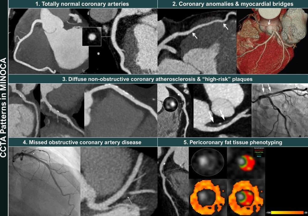
Wie soll man vorgehen, wenn bei Personen mit V. a. Angina pectoris in der CT-Koronarangiografie nichts auf eine Obstruktion hindeutet? Ein schottisches Team schlägt für solche Fälle eine Ausschlussdiagnostik mit Belastungs-EKG vor. Fehlen hier Anzeichen einer Ischämie, sind mikrovaskuläre Störungen als Ursache für die Beschwerden unwahrscheinlich.
Das Wichtigste in Kürze zu dieser Studie finden Sie am Ende des Artikels.
Bei unklaren Beschwerden in der Brust und Angina-pectoris-Verdacht kann eine CT-Untersuchung der Koronargefäße (CCTA) evtl. vorhandene Engstellen aufdecken. Allerdings gibt es viele Fälle, in denen auf diese Weise keine Obstruktion gefunden wird (ANOCA: Angina ohne obstruktive Koronararterien bzw. INOCA: Ischämie ohne obstruktive Koronararterien). In solchen Fällen könnte zum Beispiel ein Endotyp mit mikrovaskulärer Angina, vasospastischer Angina oder beidem vorliegen.
Nach Robert Sykes vom West of Scotland Heart and Lung Centre in Glasgow und seinem Team könnte hier ein einfaches Belastungs-EKG weiterhelfen.
Verdachtsdiagnose ANOCA
In ihre Studie schlossen sie 163 Personen (Durchschnittsalter 55 Jahre; Frauenanteil 63%) mit suspizierter ANOCA bzw. INOCA aus der CorCTA-Studie ein. Nach der Herz-CT, welche in 42% der Fälle keinen Hinweis auf eine koronare Atherosklerose erbracht hatte, wurden verschiedene invasive Koronarfunktionstests zur Endotypisierung durchgeführt: ein Führungsdrahttest zur Messung der Koronarflussreserve (CFR) und des mikrovaskulären Widerstands (IMR), gefolgt von einer intrakoronaren Infusion von Acetylcholin zur Messung der Vasospasmusneigung.
Das Post-CCTA-Belastungs-EKG wurde nach dem Bruce-Protokoll auf dem Laufband absolviert. Demnach waren knapp 28% der Teilnehmenden ischämisch, definiert als ST-Strecken-Senkung ≥ 0,1 mV; von den Personen ohne Hinweis auf Atherosklerose im CT betraf das jede/n dritte/n. In der Ischämiegruppe war der Wert im Rose Angina Questionnaire (RAQ), der die Stabilität der Angina misst, signifikant schlechter als in der nichtischämischen Gruppe (48,3 gegenüber 55,2).
Belastungstest als Ausschluss-Strategie
Der Ischämiestatus im Belastungs-EKG (ischämisch oder nicht ischämisch) hatte eine geringe Sensitivität (ca. 30%) und moderate Spezifität (ca. 73%), aber einen hohen negativen Vorhersagewert (NPV) für das Vorhandensein einer mikrovaskulären Dysfunktion. Im Einzelnen betrug der NPV
94,1% für die Kombination CFR < 2,5, Spasmus und IMR ≥ 25 (zehn Fälle),
92,4% für CFR < 2,5 und mikrovaskulären Spasmus (15 Fälle),
85,6% für CFR < 2,5 und IMR ≥ 25 (n = 22) und
81,4% für CFR < 2,5 allein (n = 33).
Für den alleinigen mikrovaskulären Spasmus wurde allerdings nur ein NPV von 58,5% ermittelt (n = 70).
Sykes und sein Team schlagen folgenden Algorithmus für Personen mit vermuteter Angina pectoris vor: Initial solle eine CCTA erwogen werden. Bestehe danach der Verdacht auf eine ANOCA/INOCA, könne das Belastungs-EKG zum Ausschluss mikrovaskulärer Endotypen, zur Ermittlung der Belastungskapazität und für die Prognose genutzt werden.
Das Wichtigste in Kürze
Frage: Diagnostische Genauigkeit eines Belastungs-EKG nach CT-Koronarangiografie (CCTA) zur Identifizierung bzw. zum Ausschluss einer mikrovaskulären Dysfunktion bei Angina-pectoris-Beschwerden.Antwort: Der negative prädiktive Wert (NPV) des Ischämiestatus laut EKG war hoch, der positive Vorhersagewert dagegen niedrig.Bedeutung: Mikrovaskuläre Spasmen oder eine mikrovaskuläre Angina können bei fehlenden Hinweisen auf eine Ischämie im Belastungs-EKG mit relativ hoher Sicherheit ausgeschlossen werden.Einschränkung: Keine anderen diagnostischen Tests berücksichtigt.
Alkoqolun oranizmə olan mənfi təsirləri haqqında hər birimiz eşitmişik. Lakin bu gün onun ürəyə olan ziyanlı təsiri haqqinda danışaq. Bəzi tədqiqatlar az miqdarda alkoqolun oranizmə ziyanlı olmaması haqqında qeyd etsə də, lakin bu tam elmi təsdiq olunmayıb. Bundan başqa Alokoqol qəbul edən şəxslərin, çox zaman bu təyin olunmuş az miqdarı aşmasını nəzərə alsaq zərərlər daha aydın olacaq.
Zərərli təsir həm damarlar həmdə ürəyin əzələsində baş verir. Belə ki kiçik və orta ölçülü damarlar ilk əvvəl genişlənir və arterial təzyiq aşağı enir ki, bu da özününyaxşı hiss etmə illuziyası yaradır. Lakin bir neçə saat sonar orqanizmdə kompensator olaraq damarların daralması və təzyiqin yüksəlməsi baş verir ki, bu artıq 1 sutkaya qədər davam edir. Alkoqolun təsiri bununla bitmir. Etanolun təsirindən kardiositlərin istehsalı azalır ki, bu da ürək əzələsində enerji çatmazlığı yaradır. Ürək əzələsi zədələnir, funksiyası zəifləyir, zəncirvari olaraq ürək çatmazlığı və müxtəlif ritm pozğunluqlarının yaranması başlayır. Damarlar təsir nəticəsində yüksək qan təzyiq-arterial hipertoniya, ürəyə təsir isə stenokardiya,miokard infarktı,ürək çatmazlığı,kardiomiopatiya, taxikardiya və müxtəlif ritm pozğunluğu kimi xəstəliklərin inkişafı başlayır. Davamlı qəbul edilən alkoqol isə bu xəstəliklərin dərinləşməsinə və geridönməz dəyişikliklərə gətirir. Orqanizmə tək Etanol zərər vermir. Alokoqollu içkilərin tərkibində olan müxtəlif qatqılar və efir yağları əlavə zədələnmələrə gətirir. Bu təsirlər əsasən də kustar üsulla hazırlanmış alkoqollu içkilər, viski, pivə və digərlərində daha qabarıq özünü göstərir.
Orqanizmin funksiyaların tənzilmlənməsində hormonların rolunu yəqin ki, eşitmisiz. Müxtəlif yaşlarda müxtəlif orqanların funksiyası daima Hormonların nəzarəti altında olur və onların qanda miqdarının dəyişməsi müxtəlif dəyişikliklərə səbəb olur. Lakin bugün məqsəd onların ürək funksiyasına necə təsir etməsini aydınlaşdırmaqdır. Bu qadınlarda və kişilərdə istehsal olunan hormonlara görə müxtəlif olur.
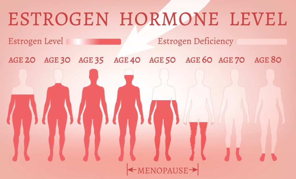
Qadınlarda bu ilk növbədə əsas hormon sayılan Esterogenlər miqdarında əks olunur.Belə ki, onlar damarları qoruyur,aretriyaların elastikliyini artırır,ateroskleroz yaranmasını əngəlləyir ki, bu da xolesterin yastıqcıqlarının formalaşmasının qarşısını alır. Bundan başqa digər əhəmiyyətli hormon olan Progesteronda təsiri əhəmiyyətli sayılır. Hormon damar divarının normal tonusda qalmasına kömək edərək ödemlərin yaranmasını qarşısını alır. Qadın orqanlarında yaranmış müxtəlif xəstəliklər ( endometrioz və yumurtalıqların polikistozu kimi ginekoloji problemlər) və Menopauza(klimaks) sonra yaranmış hormonal disfunksiya növbəti illərdə yüksək qan təzyiqi, şəkərli diabet və ürək işemik xəstəliyinin inkişafına səbəb olur. Xroniki stresin yaranması isə bəzi qadınlarda “Sınmış Ürək” kimi tanınan kardiomiopatiya yaranmasına gətirə bilər.
Kişilərdə isə əsas hormon Testesteron sayılır. Bu hormonun əsas təsiri Əzələ kütləsinin inkişafını tənzimləməkdir. Ürək də əzələ sayıldığından bu özünü daha qabarıq göstərir. Bundan başqa testesteron qanda olan “Pis xolesterinin” yəni aşağı sıxlıqlı xolesterinin qanda yüksəlməsinin qarşısını almaqla gələcəkdə damar divarında xolesterin yığılmasını nəzərəçarpan dərəcə aşağı salır.
Bunlardan başqa müxtəlif stress hallarında və reaktiv proseslər zamanı qanda kortizol və adrenalinin miqdarının artması ürək döyüntülərinin sayının artması,arterial təzyiqin yüksəlməsi, müxtəlif ritm pozulmalarının yaranması ola bilər ki, nəticədə yeni ürək xəstəliklərinin inkişafına səbəb olur.
Hormonlarınızın miqdarını tənzimləməklə Öz ürəyinizidə qorumuş olursuz!
Nə qədər qəribə səslənsədə belə fərqlər var. Dünyanın müxtəlif ölkələrinin qabaqcıl Tibbi Mərkəzlərində bu mövzuda çoxsaylı tədqiqatlar aparılmışdır və bu fərqlər həm ürəyin anatomiyasında həm də yarana biləcək xəstəliklərdə özünü göstərməkdədir. Bəs bu fərqlər hansılardır?
Ürək və damarların ölçüsü-Hər kəsin ürəyi anatomik və quruluşca eyni olsa da ,qadınlarda fərqli olaraq onun və kameralarının ölçüləri daha kiçik, damarları daha nazik olur.
Hormonlar – Əsasəndə qadınlarda baş verən hormonal pozğunluqlar (Postmenopausa yəni Klimaks zamanı) qanda Esterogenlərin və Testesteronun miqdarında baş verən dəyişiklər yüksək qan təzyiqinin, şəkərli diabet və ürək damar xəstəliklərinin artmasına səbəb olur.
Risk faktorlar– Qadınlarda və kişilərdə ciddi fərlənməkdədir. Kişilərin əsas risk faktorları – siqaret,alkoqol ,stress və pozulmuş qidalanma sayılsada qadınlarda bu erkən yaranmış arterial hipertoniya və şəkərli diabet, artıq çəki və hipodinamiya sayılır.
Xəstəliyin yaşı-Qadınlarda Ürək-damar xəstəlikləri kişilərə nisbətən 7-10 il daha gec yaşlarda üzə çıxır.Bu səbəbdən gündəlik həyatda kişilərin daha cavan yaşlarda miokard infarkt olmasını eşidirik.
Ürək Ritm pozğunluqları– qadınlarda göstərdiyimiz hormonal çatışmazlıq və kiçik arterial zədələnməsi nəticəsində müxtəlif Taxikardiya və Səyirici Aritmiya kimi ritm pozğunluqlarının daha sıx rast gəlinməsi görülür.
Simptomlar– Ürək damar hadisələrinin kəskinləşməsi-Kişilərdə döş sümüyü arxası ağrı və digər simptomların daha kəskin özünü biruzə verməsi ilə olursa, qadınlarda daha çox kəskin ürəkdöyünmə, ürəkbulanma,qusma,soyuq tər, yüksək qan təzyiqi kimi simptomlarla önə çıxır.
Yüksək qan təzyiqi olan şəxslər hər dəfə dərman qəbul etməlidirmi? Xeyr – əsasən də bu cür Hipertenziya ümumi yorğunluq,stress, iş gərginliyi və s qeyri orqanik xəstəliklər fonunda yaranarsa. Sizə qan təzyiqinin dərmansız normaya salınmasının 10 Yolunu göstərim.
Artıq çəkidən azad olun – Qan təzyiqinin bədən çəkisi ilə bir başa əlaqəli olması artıq çoxdan məlumdur. Aparılmış tədqiqatlar göstərib ki, bədən çəkisinin hər 1 kg endirilməsi qan təziyinində 1 mmHg enməsinə səbəb olur. Bundan başqa bel çevrəsinin ölçüsüdə çox vacib parametr sayılır. Belə ki, kişilərdə bu ölçü 102 sm, qadınlarda isə 89 sm yuxarı olması Yüksək qan təzyiqi riskinə işarədir.
Mütəmadi fiziki aktivlik– qan təzyiqinin ən azı 5-8 mmhg aşağı enməsinə səbəb olur. Fiziki aktivlik dedikdə həftədə ən azı 2-3 dəfə 30-40 dəq müddətdə olmaqla yüngül atletik idmanlar(gəzinti, yüngül qaçış,velosiped sürmək,üzmək, rəqs etmək) nəzərdə tutulur.
Sağlam Qidalar yeyin– gündəlik qida rasionuzda daha çox meyvə tərəvəz və az yağlı qidalar olsun.Aralıq dənizi dietası və qəbul edilən qidaların kaliumla bol olması qan təzyiqinin 11 mmHg qədər enməsinə gətirir.
Xörək duzu qəbulunu azaldın– qidalarınız az duzlu olması( gündəlik Na qəbulunun 1500 mg və daha aşağı olması) qan təzyiqinin 5-6 mmHg aşağı endirir. Bunun üçün qəbul etdiyiniz hər qidanın tərkibində Na və ya xörək duzunun miqdarına nəzarət edin və qəbul etdiyiniz yeməklərin və salatları duzsuz yeyin.
5. Alkoqol azaldın– Gündəlik qəbulun azaldılması qan təzyiqinin ən azı 4-5 mmHg enməsinə səbəb olur. Artıq qəbul edilmiş içki damarlarda uzunmüddətli daralmaya,qanın laxtalanmasına və ürək ritminin pozulmasına gətirirki bu da Yüksək qan təzyiqilə birbaşa əlaqəli olur.
6. Tütün istifadəsi dayandırın– bu həm qan təzyiqinin enməsinə həm də gələcəkdə ürək və beyin-damar xəstəliklərinin ,Xroniki ağciyər xəstəliyi kimi halların yaranmasını azaldır.
7. Gecə yuxunuzu tənzimləyin– hər gecə yuxusu 7-9 saat olması yetkin şəxslər üçün normal sayılır. Yuxusuzluq qan təzyiqinin yüksəlməsinin əsas faktorlarından sayılır. Bunun qarşısını almaq üçün çalışın hər gün eyni vaxtlar yatın,yataq otağınızın havası optimal(21-22 C),qaranlıq,sakit, əlavə elektronik vasitələr olmayan (TV,Mobil telefon və kompyuter) bir məkan olsun.
8. Stressdən uzaq olun– uzun müddətli və çətin müalicəyə tabe olan Arterial Hipertenziyaların əsas səbəblərindən sayılır.Bunları yaradan səbəblərdən mümkün qədər uzaq qaçın.Gündəlik hobbi və aktivitələrə daha çox vaxt ayırın.
9. Qanda Xolesterol və Şəkərin miqdarını kontrolda saxlayın– bunun üçün ildə 1 dəfə qan təhlilləri görməyinizi Sizin gələcək risklərinizin qarşısını almağa daha çox kömək edər. Yüksək Şəkər və Xolesterol yüksək qan təzyiqinin dayanıqlı olmasına zəmin yaradır.
10.Qan təzyiqinizi özünüz ölçün– evinizdə və ya iş yerində təzyiqi ölçmək üçün Cihaz olsun. Narahat olduğunuz vaxtlar günün müxtəlif vaxtlarında təzyiqi ölçməklə öz həkimizi düzgün məlumatlandıra bilərsiz .


 ,Naomi Allen1 2,Ben Lacey1,Elizabeth Soilleux3,Thomas Littlejohns1