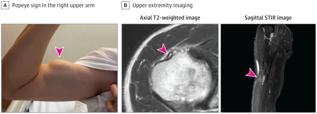
Popeye deformasiyası nədir? Popeye deformasiyası, cizgi filmi qəhrəmanı Popeye-in həddindən artıq böyük bisepsinə bənzəyən şişkin biseps əzələsi ilə xarakterizə olunur. Bu vəziyyət adətən biseps əzələsini çiynə birləşdirən biseps tendonunun uzun başındakı yırtıqdan yaranır. Lakin bəzən bu hal sistem xəstəlikləri zamanıda yarana bilir. 2022 ci ildi Avropa Kardioloji Cəmiyyətinin aylıq dərgisində yayımlanmış məqalədə bir neçə Yapon və İspan alimləri bu haqda çox maraqlı araşdırma aparmışlar. Bu araşdırmaların qısa icmalı original dildə təqdim olunur.
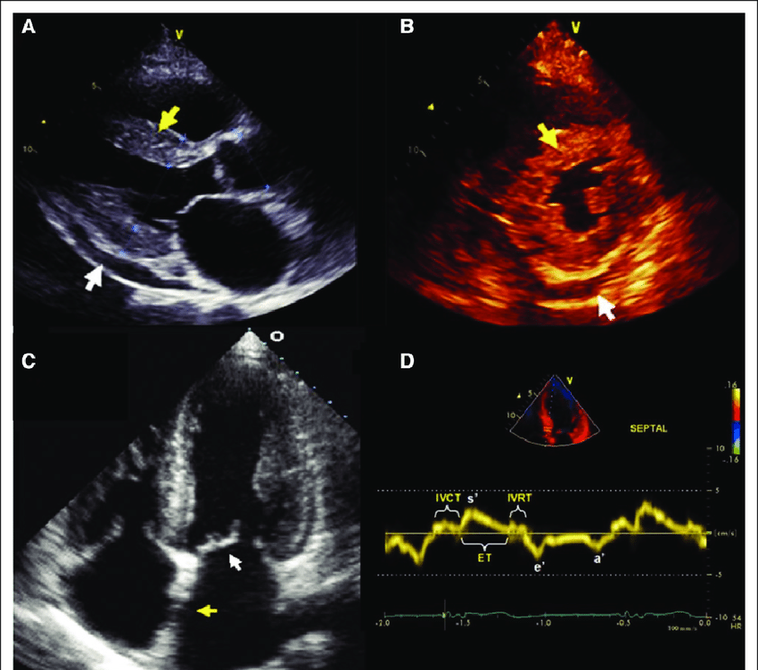
A 76-year-old man with a history of cervical open-door laminoplasty for ossification of the posterior longitudinal ligament presented to our hospital with a symptom of exertional dyspnoea. A 12-lead electrocardiogram revealed I-degree atrioventricular block (PQ interval 262 ms), left axis deviation, and complete right bundle branch block (Panel A). A transthoracic echocardiogram indicated left ventricular hypertrophy with a maximum wall thickness of 17 mm and a left ventricular ejection fraction of 61% (Panel B1). Strain echocardiogram using speckled tracking showed marked diminution of global longitudinal strain with apical sparing (Panel B2). Physical examination found subconjunctival hemorrhage in right eye, several purpuric lesions in upper extremities, and a large bulge on his left upper arm when he flexed his arm, so-called Popeye’s sign, indicating a hallmark of biceps tendon rupture (see Supplementary material online, Video S1; Panel C; arrow). A monoclonal component was not detected by serum and urine immune-electrophoresis and free light chain assays. 99mTc-labelled bone scintigraphy showed grade 3 myocardial uptake (Panel D). Eventually, endomyocardial biopsy and genetic sequencing analysis of the transthyretin gene confirmed the patient as a wild-type transthyretin amyloidosis (Panel E). Popeye’s sign is uncommon, but pivotal to diagnose transthyretin amyloidosis.
Spain Research doünload PDF content A 69-year-old man presented to our cardiology clinic troubled by breathlessness on exertion. He had a history of hypertension, atrial fibrillation, and bilateral carpal tunnel syndrome. Physical examination showed jugular venous distension, bilateral ankle oedema, and a bunching of the right biceps when he flexed his arm—so called Popeye’s sign—indicating rupture of the proximal biceps tendon (figure and video); a rupture was confirmed by an ultrasound scan. Serum N-terminal-pro-B-type natriuretic peptide was 7088 pg/mL (normal range for patients aged 0–74 years is less than 125 pg/mL). A 12-lead electrocardiogram showed low voltage in the limb leads, as well as a pseudo-infarct pattern in the precordial ones (appendix). A transthoracic echocardiogram showed left ventricular hypertrophy with a maximum wall thickness of 20 mm and a left ventricular ejection fraction (LVEF) of 40%. Cardiac MRI showed signs of myocardial infiltration. 99mTechnetium-3,3-diphosphono-1,2-propanodicarboxylic acid (99mTc-DPD) scintigraphy showed grade 3 myocardial uptake (figure). Serum and urine immunoelectrophoresis and free light chain assays ruled out a monoclonal component. Genetic sequencing analysis of the transthyretin gene found no mutations. Tying all the findings together, a diagnosis of heart failure secondary to wild-type transthyretin amyloid (ATTRwt) cardiomyopathy was made. The patient was treated with a combination of diuretics—namely, furosemide 80 mg twice a day, hydrochlorothiazide 12・5 mg once a day, and spironolactone 100 mg once a day—but he did not respond well. He remained symptomatic with advanced functional impairment and persistent signs of systemic venous congestion. ATTRwt, previously known as senile systemic amyloidosis, is a disease that typically affects the heart and tendons of elderly patients. A history of bilateral carpal tunnel syndrome or, less frequently, spontaneous tendon rupture in a patient with heart failure and preserved or mildly depressed LVEF should raise clinical suspicion of the condition. A definitive diagnosis of ATTRwt cardiomyopathy can be confirmed non-invasively with a positive finding on 99mTc-DPD scintigraphy, together with the absence of a monoclonal component in serum and urine samples, and a normal genetic sequencing analysis of the transthyretin gene. Current therapy for patients with ATTRwt cardiomyopathy is limited, with diuretics for relief of cardiac congestion being the mainstay of treatment. However, a recent randomised, placebo controlled trial, published in September 2018, showed a significant reduction in the risk of death and hospitalisation from cardiovascular causes in patients with ATTRwt cardiomyopathy treated with tafamidis, a transthyretin stabilising agent. Tafamidis is expected to be available for clinical use in the near future.
Wie soll man vorgehen, wenn bei Personen mit V. a. Angina pectoris in der CT-Koronarangiografie nichts auf eine Obstruktion hindeutet? Ein schottisches Team schlägt für solche Fälle eine Ausschlussdiagnostik mit Belastungs-EKG vor. Fehlen hier Anzeichen einer Ischämie, sind mikrovaskuläre Störungen als Ursache für die Beschwerden unwahrscheinlich.
Das Wichtigste in Kürze zu dieser Studie finden Sie am Ende des Artikels.
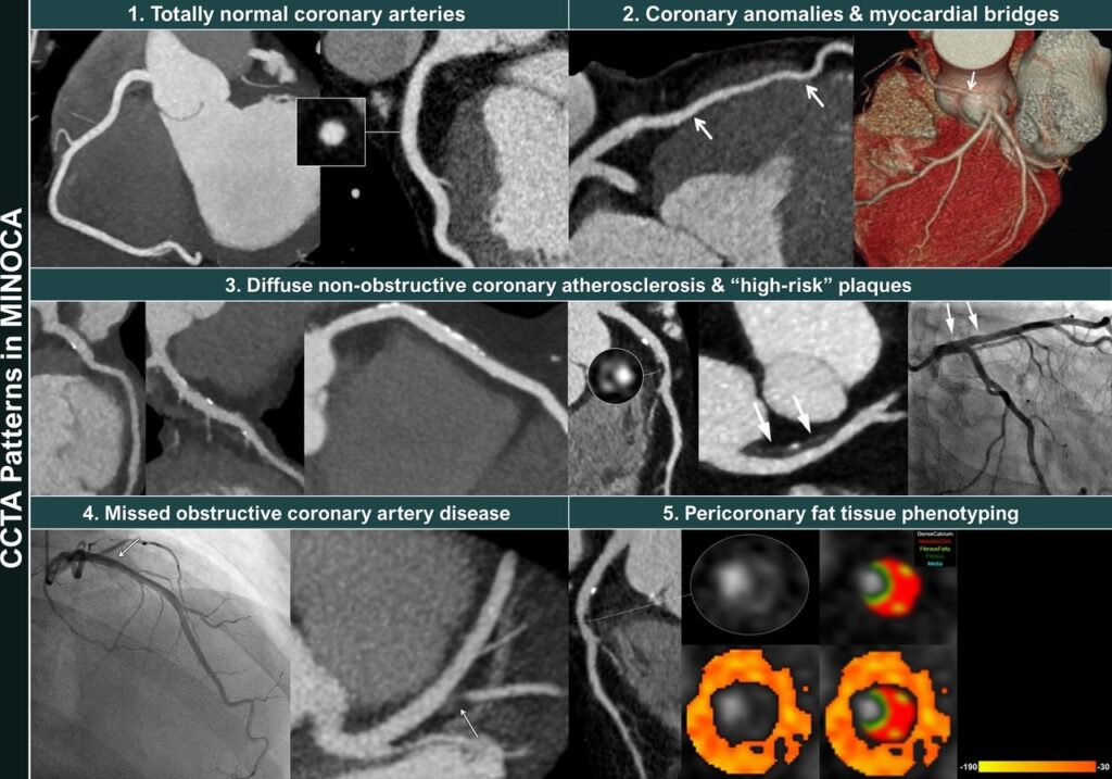
Bei unklaren Beschwerden in der Brust und Angina-pectoris-Verdacht kann eine CT-Untersuchung der Koronargefäße (CCTA) evtl. vorhandene Engstellen aufdecken. Allerdings gibt es viele Fälle, in denen auf diese Weise keine Obstruktion gefunden wird (ANOCA: Angina ohne obstruktive Koronararterien bzw. INOCA: Ischämie ohne obstruktive Koronararterien). In solchen Fällen könnte zum Beispiel ein Endotyp mit mikrovaskulärer Angina, vasospastischer Angina oder beidem vorliegen.
Nach Robert Sykes vom West of Scotland Heart and Lung Centre in Glasgow und seinem Team könnte hier ein einfaches Belastungs-EKG weiterhelfen.
Verdachtsdiagnose ANOCA
In ihre Studie schlossen sie 163 Personen (Durchschnittsalter 55 Jahre; Frauenanteil 63%) mit suspizierter ANOCA bzw. INOCA aus der CorCTA-Studie ein. Nach der Herz-CT, welche in 42% der Fälle keinen Hinweis auf eine koronare Atherosklerose erbracht hatte, wurden verschiedene invasive Koronarfunktionstests zur Endotypisierung durchgeführt: ein Führungsdrahttest zur Messung der Koronarflussreserve (CFR) und des mikrovaskulären Widerstands (IMR), gefolgt von einer intrakoronaren Infusion von Acetylcholin zur Messung der Vasospasmusneigung.
Das Post-CCTA-Belastungs-EKG wurde nach dem Bruce-Protokoll auf dem Laufband absolviert. Demnach waren knapp 28% der Teilnehmenden ischämisch, definiert als ST-Strecken-Senkung ≥ 0,1 mV; von den Personen ohne Hinweis auf Atherosklerose im CT betraf das jede/n dritte/n. In der Ischämiegruppe war der Wert im Rose Angina Questionnaire (RAQ), der die Stabilität der Angina misst, signifikant schlechter als in der nichtischämischen Gruppe (48,3 gegenüber 55,2).
Belastungstest als Ausschluss-Strategie
Der Ischämiestatus im Belastungs-EKG (ischämisch oder nicht ischämisch) hatte eine geringe Sensitivität (ca. 30%) und moderate Spezifität (ca. 73%), aber einen hohen negativen Vorhersagewert (NPV) für das Vorhandensein einer mikrovaskulären Dysfunktion. Im Einzelnen betrug der NPV
94,1% für die Kombination CFR < 2,5, Spasmus und IMR ≥ 25 (zehn Fälle),
92,4% für CFR < 2,5 und mikrovaskulären Spasmus (15 Fälle),
85,6% für CFR < 2,5 und IMR ≥ 25 (n = 22) und
81,4% für CFR < 2,5 allein (n = 33).
Für den alleinigen mikrovaskulären Spasmus wurde allerdings nur ein NPV von 58,5% ermittelt (n = 70).
Sykes und sein Team schlagen folgenden Algorithmus für Personen mit vermuteter Angina pectoris vor: Initial solle eine CCTA erwogen werden. Bestehe danach der Verdacht auf eine ANOCA/INOCA, könne das Belastungs-EKG zum Ausschluss mikrovaskulärer Endotypen, zur Ermittlung der Belastungskapazität und für die Prognose genutzt werden.
Das Wichtigste in Kürze
Frage: Diagnostische Genauigkeit eines Belastungs-EKG nach CT-Koronarangiografie (CCTA) zur Identifizierung bzw. zum Ausschluss einer mikrovaskulären Dysfunktion bei Angina-pectoris-Beschwerden.Antwort: Der negative prädiktive Wert (NPV) des Ischämiestatus laut EKG war hoch, der positive Vorhersagewert dagegen niedrig.Bedeutung: Mikrovaskuläre Spasmen oder eine mikrovaskuläre Angina können bei fehlenden Hinweisen auf eine Ischämie im Belastungs-EKG mit relativ hoher Sicherheit ausgeschlossen werden.Einschränkung: Keine anderen diagnostischen Tests berücksichtigt.
2023 ACC Expert Consensus Decision Pathway on Comprehensive Multidisciplinary Care for the Patient With Cardiac Amyloidosis: A Report of the American College of Cardiology Solution Set Oversight Committee. J Am Coll Cardiol 2023;Jan 23
The following are key points to remember from a new Expert Consensus Decision Pathway document on comprehensive multidisciplinary care for the patient with cardiac amyloidosis:
Amyloid cardiomyopathy is caused by misfolding of (i) monoclonal immunoglobulin light chain produced in bone marrow plasma cell disorders called AL, or (ii) transthyretin (TTR) protein called ATTR. ATTR-cardiomyopathy (ATTR-CM) can occur in the context of genetically normal protein (wild type or ATTRwt-CM) or due to genetic mutations (most commonly isoleucine substitution for valine at position 122), rendering the protein abnormal (ATTRv-CM).
Since amyloid fibrils can deposit in multiple organs, multidisciplinary care is a requisite.
Cardiac clues to diagnosis of amyloidosis includes increased left ventricular hypertrophy in the absence of hypertension or valvular heart disease, heart failure symptoms, diastolic dysfunction, atrial fibrillation, conduction system disease, and elevated cardiac biomarkers. Extracardiac manifestations include carpal tunnel syndrome, spinal stenosis, hip or knee replacement, prior shoulder surgery, proteinuria, and peripheral or autonomic neuropathy causing orthostatic hypotension. Pathognomonic extracardiac findings for AL include macroglossia, periorbital purpura, and acquired factor X deficiency. Findings unique to ATTR are spontaneous biceps rupture and spinal stenosis.
Low voltage electrocardiography and presence of hypertrophy on echocardiography is only present in 30% of amyloid patients. Other echo findings include left ventricular hypertrophy, atrioventricular valve/right ventricular free wall/interatrial septum thickening, diastolic dysfunction, biatrial enlargement, and decreased global longitudinal strain with apical sparing.
Cardiac magnetic resonance imaging can help differentiate amyloidosis from other infiltrative diseases. Findings include extracellular volume expansion and diffuse late gadolinium enhancement.
The first step in identifying type of amyloidosis is a monoclonal protein screen involving: serum free light chain assay, serum and urine immunofixation electrophoresis. If all are negative, AL has been ruled out. If any are positive, the next step is biopsy of the involved organ with mass spectrometry to confirm AL deposition. A negative fat pad biopsy does not rule out AL or ATTR, and biopsy of the involved organ (heart or kidneys) should be considered.
A serum/urine protein electrophoresis should not be used to rule out monoclonal protein due to lower accuracy relative to immunofixation. In chronic kidney disease, elevated serum free light chain ratios of K/L are common but with a normal serum and urine immunofixation electrophoresis.
If AL has been ruled out, a technetium pyrophosphate scan can be used to diagnose ATTR-CM. Genetic testing is warranted to distinguish between ATTRwt-CM or ATTRv-CM.
Tafamidis is a TTR stabilizer and is the only Food and Drug Administration approved medication available for all ATTR-CM. It delays disease progression but does not result in regression, and in trials, reduced all-cause mortality and cardiovascular hospitalizations. It has minimal side effects but has a high cost, needing copay assistance programs for patients.
Alternatives to tafamidis include diflunisal, also a TTR stabilizer, which is less effective but significantly cheaper. It is a nonsteroidal anti-inflammatory drug and should be avoided in chronic kidney disease, decompensated heart failure, and gastrointestinal (GI) bleeding.
Beta-blockers should be used with caution and may worsen outcomes. Angiotensin inhibitors may be poorly tolerated due to orthostatic hypotension. Retrospective analysis of trials suggests a beneficial effect of spironolactone. There is no evidence to guide use of SGLT-2 inhibitors in amyloidosis.
If atrial fibrillation is present, anticoagulation is recommended regardless of CHA2DS2-VASc score, and prior to cardioversion, a transesophageal echocardiogram should always be performed regardless of anticoagulation status due to high risk for intracardiac thrombus.
There should be close monitoring for conduction disease and ventricular arrhythmias, both being very common in amyloid cardiomyopathy. However, defibrillators have not consistently demonstrated improved survival for amyloidosis and hence should be considered based on standard heart failure guidelines for amyloid patients.
If aortic stenosis is present with cardiac amyloidosis, aortic valve replacement (AVR) may help improve symptoms and referral for transcatheter AVR (TAVR) should be considered.
Emerging therapies not yet approved for cardiac amyloidosis include TTR silencers such as patisiran or vutisiran (approved for ATTRv polyneuropathy) and inotersen. These are currently only approved for amyloid polyneuropathy. Data on green tea derivatives are lacking and not recommended.
GI mucosa involvement can cause protein losing enteropathy, GI dysmotility, abdominal pain, constipation, or diarrhea and bleeding. Treatment is supportive and should include collaboration with a GI specialist.
Treatment for multiple myeloma with AL amyloidosis includes either high-dose melphalan with stem cell transplant daratumumab with cyclophosphamide, bortezomib, and dexamethasone. For AL-CM, a stem cell transplant followed by cardiac transplant would be ideal, but in some cases, sequential heart transplant followed by stem cell transplantation may be considered.
Kidney involvement is very common with AL and ATTR amyloidosis. Treatment is supportive and includes low salt diet, use of diuretics for fluid overload, and angiotensin antagonists for proteinuria.
The American Diabetes Association (ADA) “Standards of Care in Diabetes” includes the ADA’s current clinical practice recommendations and is intended to provide the components of diabetes care, general treatment goals and guidelines, and tools to evaluate quality of care. Members of the ADA Professional Practice Committee, an interprofessional expert committee, are responsible for updating the Standards of Care annually, or more frequently as warranted. For a detailed description of ADA standards, statements, and reports, as well as the evidence-grading system for ADA’s clinical practice recommendations and a full list of Professional Practice Committee members, please refer to Introduction and Methodology. Readers who wish to comment on the Standards of Care are invited to do so at professional.diabetes.org/SOC.
Chronic Kidney Disease
Screening
Recommendations
11.1a At least annually, urinary albumin (e.g., spot urinary albumin-to-creatinine ratio [UACR]) and estimated glomerular filtration rate [eGFR] should be assessed in people with type 1 diabetes with duration of ≥5 years and in all people with type 2 diabetes regardless of treatment. B
11.1b In people with established chronic kidney disease (CKD), urinary albumin (e.g., spot UACR) and eGFR should be monitored 1–4 times per year depending on the stage of the kidney disease
11.2 Optimize glucose management to reduce the risk or slow the progression of CKD. A
11.3 Optimize blood pressure control and reduce blood pressure variability to reduce the risk or slow the progression of CKD and reduce cardiovascular risk. A
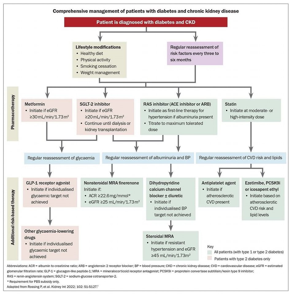
11.4a In nonpregnant people with diabetes and hypertension, either an ACE inhibitor or an angiotensin receptor blocker (ARB) is recommended for those with moderately increased albuminuria (UACR 30–299 mg/g creatinine) B and is strongly recommended for those with severely increased albuminuria (UACR ≥300 mg/g creatinine) and/or eGFR <60 mL/min/1.73 m2 to prevent the progression of kidney disease and reduce cardiovascular events. A
11.4b Periodically monitor for increased serum creatinine and potassium levels when ACE inhibitors, ARBs, and mineralocorticoid receptor antagonists are used, or for hypokalemia when diuretics are used. B
11.4c An ACE inhibitor or an ARB is not recommended for the primary prevention of CKD in people with diabetes who have normal blood pressure, normal UACR (<30 mg/g creatinine), and normal eGFR. A
11.4d Do not discontinue renin-angiotensin system blockade for mild to moderate increases in serum creatinine (≤30%) in the absence of signs of extracellular fluid volume depletion. A
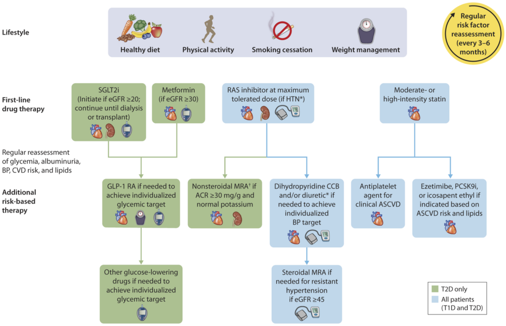
11.5a For people with type 2 diabetes and CKD, use of a sodium–glucose cotransporter 2 (SGLT2) inhibitor is recommended to reduce CKD progression and cardiovascular events in individuals with eGFR ≥20 mL/min/1.73 m2 and urinary albumin ≥200 mg/g creatinine. A
11.5b For people with type 2 diabetes and CKD, use of an SGLT2 inhibitor is recommended to reduce CKD progression and cardiovascular events in individuals with eGFR ≥20 mL/min/1.73 m2 and urinary albumin ranging from normal to 200 mg/g creatinine. B
11.5c For cardiovascular risk reduction in people with type 2 diabetes and CKD, consider use of an SGLT2 inhibitor (if eGFR is ≥20 mL/min/1.73 m2), a glucagon-like peptide 1 agonist, or a nonsteroidal mineralocorticoid receptor antagonist (if eGFR is ≥25 mL/min/1.73 m2). A
11.5d As people with CKD and albuminuria are at increased risk for cardiovascular events and CKD progression, a nonsteroidal mineralocorticoid receptor antagonist that has been shown to be effective in clinical trials is recommended to reduce cardiovascular events and CKD progression (if eGFR is ≥25 mL/min/1.73 m2). Potassium levels should be monitored. A
11.6 In people with CKD who have ≥300 mg/g urinary albumin, a reduction of 30% or greater in mg/g urinary albumin is recommended to slow CKD progression. C
11.7 For people with non–dialysis-dependent stage G3 or higher CKD, dietary protein intake should be aimed to a target level of 0.8 g/kg body weight per day. A For individuals on dialysis, 1.0–1.2 g/kg/day of dietary protein intake should be considered since protein energy wasting is a major problem in some individuals on dialysis. B
11.8 Individuals should be referred for evaluation by a nephrologist if they have continuously increasing urinary albumin levels and/or continuously decreasing eGFR and/or if the eGFR is <30 mL/min/1.73 m2. A
11.9 Promptly refer to a nephrologist for uncertainty about the etiology of kidney disease, difficult management issues, and rapidly progressing kidney disease. B
Marie B. Demay (Chair), Anastassios G. Pittas (Co-Chair), Daniel D. Bikle, Dima L. Diab, Mairead E. Kiely, Marise Lazaretti-Castro, Paul Lips, Deborah M. Mitchell, M. Hassan Murad, Shelley Powers, Sudhaker D. Rao, Robert Scragg, John A. Tayek, Amy M. Valent, Judith M. E. Walsh, Christopher R. McCartney
The 2024 guideline on vitamin D for the prevention of disease:
Essential Points
Numerous studies demonstrate an association between serum concentrations of 25-hydroxyvitamin D (25[OH]D) and a variety of common disorders including musculoskeletal, metabolic, cardiovascular, malignant, autoimmune, and infectious diseases. This has led to widespread supplementation with vitamin D supplementation and increased laboratory testing for 25(OH)D in the general population.
The benefit-risk ratio of this increase in vitamin D supplementation is not clear, and the optimal vitamin D intake and serum 25(OH)D concentrations for disease prevention remain uncertain.
This guideline offers clinical guidelines for the use of vitamin D to lower the risk of disease in individuals without established indications for vitamin D treatment or 25(OH)D testing.
List of Recommendation
Question 1.Should empiric vitamin D supplementation vs no empiric vitamin D supplementation be used for children and adolescents (ages 1-18 years)?
Recommendation 1
In children and adolescents ages 1-18 years, we suggest empiric vitamin D supplementation to prevent nutritional rickets and potentially lower the risk of respiratory tract infections. (2 | ⊕⊕OO)
Empiric vitamin D may include daily intake of fortified foods, vitamin formulations that contain vitamin D and/or daily intake of a vitamin D supplement (pill or drops).
In the clinical trials included in the SR, with respect to respiratory tract infections in children, vitamin D doses ranged from 300 to 2000 IU (7.5 to 50 μg) daily equivalent. The estimated weighted average was approximately 1200 IU (30 μg) per day.
Question 2. Should empiric vitamin D supplementation vs no empiric vitamin D supplementation be used for nonpregnant adults <50 years of age?
Recommendation 2 In the general adult population younger than age 50 years, we suggest against empiric vitamin D supplementation. (2 | ⊕◯◯◯)
This recommendation relates to empiric vitamin D supplementation that exceeds the DRIs established by the IOM. Adults in this age group should follow the Recommended Daily Allowance established by the IOM (600 IU [15 µg] daily).
Question 3.Should vitamin D supplementation vs no vitamin D supplementation be used for nonpregnant adults <50 years of age only when 25(OH)D levels are below a threshold?
Recommendation 3
In the general adult population younger than age 50 years, we suggest against routine 25(OH)D testing. (2 | ⊕OOO)
In this population, 25(OH)D levels that provide outcome-specific benefits have not been established in clinical trials.
The panel suggests against (a) routine screening for a 25(OH)D level to guide decision-making (i.e., vitamin D vs no vitamin D) and (b) routine follow-up testing for 25(OH)D level to guide vitamin D dosing.
This recommendation relates to generally healthy adults who do not otherwise have established indications for 25(OH)D testing (e.g., hypocalcemia).
Question 4. Should empiric vitamin D supplementation vs no empiric vitamin D supplementation be used for adults aged 50-74 years?
Recommendation 4
In the general population ages 50 to 74 years, we suggest against routine vitamin D supplementation. (2 | ⊕⊕⊕O)
This recommendation relates to empiric vitamin D supplementation that exceeds the DRIs established by the IOM. Adults in this age group should follow the Recommended Daily Allowance established by the IOM (600 IU [15 μg] daily for those aged 50 to 70 years; 800 IU [20 μg] daily for those older than 70 years).
Question 5. Should vitamin D supplementation vs no vitamin D supplementation be used for adults aged 50-74 years only when 25(OH)D levels are below a threshold?
Recommendation 5
In the general population ages 50 to 74 years, we suggest against routine 25(OH)D testing. (2 | ⊕OOO)
In this population, 25(OH)D levels that provide outcome-specific benefits have not been established in clinical trials.
The panel suggests against (a) routine screening for a 25(OH)D level to guide decision-making (i.e., vitamin D vs no vitamin D) and (b) routine follow-up testing for 25(OH)D level to guide vitamin D dosing.
This recommendation relates to generally healthy adults who do not otherwise have established indications for 25(OH)D testing (e.g., hypocalcemia).
Question 6. Should empiric vitamin D supplementation vs no empiric vitamin D supplementation be used by adults ages >75 years?
Recommendation 6
In the general population ages 75 years and older, we suggest empiric vitamin D supplementation because of the potential to lower the risk of mortality. (2 | ⊕⊕⊕O)
Empiric vitamin D may include daily intake of fortified foods, vitamin formulations that contain vitamin D and/or daily intake of a vitamin D supplement.
For empiric supplementation, daily, lower-dose vitamin D is preferred over non-daily, higher doses.
In the clinical trials included in the SR that reported on the mortality outcome, vitamin D dose ranged from 400 to 3333 IU [10 to 83 μg] daily equivalent. The estimated weighted average was average was approximately 900 IU (23 μg) daily. Participants in many trials were allowed to remain on their routine supplements, including up to 800 IU (20 μg) of vitamin D daily.
Question 7. Should vitamin D supplementation vs no vitamin D supplementation be used by adults ages >75 years only when 25(OH)D levels are below a threshold?
Recommendation 7
In the general population ages 75 years and older, we suggest against routine testing for 25(OH)D levels. (2 | ⊕OOO)
In this population, 25(OH)D thresholds that provide outcome-specific benefits have not been established in clinical trials.
The panel suggests against (a) routine screening for a 25(OH)D level to guide decision-making (i.e., vitamin D vs no vitamin D) and (b) routine follow-up testing for 25(OH)D level to guide vitamin D dosing.
This recommendation relates to generally healthy adults who do not otherwise have established indications for 25(OH)D testing (e.g., hypocalcemia).
Question 8. Should empiric vitamin D supplementation vs no empiric vitamin D supplementation be used during pregnancy?
Recommendation 8
We suggest empiric vitamin D supplementation during pregnancy, given its potential to lower risk of preeclampsia, intra-uterine mortality, preterm birth, small for gestational age birth, and neonatal mortality. (2 | ⊕⊕OO)
This recommendation is based on evidence from trials conducted in healthy individuals during pregnancy.
Empiric vitamin D may include daily intake of fortified foods, prenatal vitamin formulations that contain vitamin D, and/or a vitamin D supplement (pills or drops).
In the clinical trials included in the SR, the vitamin D doses ranged from 600 to 5000 IU (15 -125 μg) daily equivalent, usually provided daily or weekly. The estimated weighted average was approximately 2500 IU (63 μg) per day.
Question 9. Should vitamin D supplementation vs no vitamin D supplementation be used during pregnancy only when 25(OH)D levels are below a threshold?
Recommendation 9
During pregnancy, we suggest against routine 25(OH)D testing. (2 | ⊕OOO)
In this population, 25(OH)D levels that provide pregnancy outcome-specific benefits have not been established in clinical trials.
The panel suggests against (a) routine screening for a 25(OH)D level to guide decision-making (ie, vitamin D vs no vitamin D) and (b) routine follow-up testing for 25(OH)D level to guide vitamin D dosing.
This recommendation relates to generally healthy pregnant individuals who do not otherwise have established indications for 25(OH)D testing (e.g., hypocalcemia).
Question 10. Should empiric vitamin D supplementation vs no empiric vitamin D supplementation be used for adults with prediabetes (by glycemic criteria)?
Recommendation 10
For adults with high-risk prediabetes, in addition to lifestyle modification, we suggest empiric vitamin D supplementation to reduce the risk of progression to diabetes. (2 | ⊕⊕⊕O)
Lifestyle modification must be a routine management component for adults with prediabetes.
The clinical trials informing this recommendation primarily related to adults with high-risk prediabetes, identified as meeting two or three American Diabetes Association glycemia criteria (fasting glucose, HbA1c, 2-hour glucose after a 75-gram oral glucose challenge) for prediabetes and those with impaired glucose tolerance.
In the clinical trials included in the SR, the vitamin D doses ranged from 842 to 7543 IU (21 to 189 μg) daily equivalent. The estimated weighted average was approximately 3500 IU (88 μg) per day. Participants in some trials were allowed to remain on their routine supplements, including up to 1000 IU (25 μg) of vitamin D daily.
Question 11. Should a daily, lower-dose vitamin D vs. non-daily (i.e., intermittent), higher-dose vitamin D be used for nonpregnant people for whom vitamin D treatment is indicated?
Recommendation 11
In adults ages 50 years and older who have indications for vitamin D supplementation or treatment, we suggest daily, lower-dose vitamin D instead of non-daily, higher-dose vitamin D. (2 | ⊕⊕OO)
The panel did not identify evidence related to individuals younger than age 50 years.
Question 12. Should screening with a 25(OH)D test (with vitamin D supplementation/treatment only if below a threshold) vs no screening with a 25(OH)D test be used for healthy adults?
Recommendation 12
In healthy adults, we suggest against routine screening for 25(OH)D levels. (2 | ⊕OOO)
In healthy adults, 25(OH)D levels that provide outcome-specific benefits have not been established in clinical trials.
This recommendation relates to adults who do not otherwise have established indications for testing with 25(OH)D levels (e.g., hypocalcemia).
Question 13. Should screening with a 25(OH)D test (with vitamin D supplementation/treatment only if below a threshold) vs no screening with a 25(OH)D test be used for adults with dark complexion?
Recommendation 13
In adults with dark complexion, we suggest against routine screening for 25(OH)D levels. (2 | ⊕OOO)
This recommendation relates to generally healthy adults with dark complexion who do not otherwise have established indications for 25(OH)D testing (e.g., hypocalcemia).
The panel did not identify any clinical trials that related clinical outcomes to skin complexion per se. A secondary analysis did not clearly suggest net benefit with vitamin D in those who self-identify as Black. The panel recognized that self-identified race is an inaccurate and otherwise problematic proxy for dark complexion.
Question 14. Should screening with a 25(OH)D test (with vitamin D supplementation/treatment only if below a threshold) vs no screening with a 25(OH)D test be used for adults with obesity?
Recommendation 14
In adults with obesity, we suggest against routine screening for 25(OH)D levels. (2 | ⊕OOO)
In adults with obesity, 25(OH)D thresholds that provide outcome-specific benefits have not been established in clinical trials.
This recommendation relates to generally healthy adults with obesity who do not otherwise have established indications for 25(OH)D testing (e.g., hypocalcemia).
Dyslipidemia is common in the general population and increases the risk of cardiovascular disease. Treatment of dyslipidemia is effective in decreasing morbidity from cardiovascular disease. Because the liver is the primary source of cholesterol and other lipids in the body, medications for dyslipidemia, such as statins, target genes in the liver. Furthermore, the liver plays a role in the metabolism of many drugs, including those that are used to treat dyslipidemia. It is not surprising, therefore, that many practitioners are hesitant to prescribe medicines to treat dyslipidemia in the setting of liver disease. This update is aimed at summarizing current understanding of the safety of treating patients who have various liver diseases and dyslipidemia with lipid-lowering drugs (Table 1).
Table 1.
Best Practice Advice: Treatment of Dyslipidemia in Liver Disease
DILI
BPA #1 Statins often (3%) cause benign elevations in serum ALT or AST levels and should not be considered contraindicated in patients with liver disease.
BPA #2 Liver biochemical tests are recommended before starting a statin but do not need to be checked routinely while statins are taken unless clinically significant side effects develop.
BPA #3 Increases in serum ALT or AST levels to >3 times ULN with evidence of cholestasis (bilirubin >2 times ULN) (in the absence of biliary obstruction) after statins are started generally require that the statin be stopped. A work-up for DILI should include testing for the presence of other underlying causes of liver disease or other medications that may have precipitated the reaction besides or in addition to the statin.
BPA #4 If DILI or ALF occurs in a patient taking a statin, other statins should be avoided in that patient.
BPA #5 DILI and ALF caused by statins are rare (1 in 100,000 and 1 in 1,000,000, respectively), so fear of developing these effects should not be used to justify avoidance of statins when an individual may benefit from them.
BPA #6 Statins are contraindicated in patients with ALF because of the patients’ poor prognosis.
BPA #7 Other lipid-lowering medications, such as niacin, ezetimibe, or fibrates, may cause DILI, but such instances are exceedingly rare and should not prevent starting these medications in a patient who may benefit from them.
NAFLD
BPA #8 Although NAFLD and NASH are not considered traditional risk factors for cardiovascular disease, they are associated with dyslipidemia. The 2013 ACC/AHA guidelines should be used to assess cardiovascular risk in patients with NAFLD and to guide the need for lipid-lowering pharmacotherapy.
BPA #9 Statins, ezetimibe, omega-3 fatty acids, and fibrates are safe and well tolerated in the setting of NAFLD and NASH.
BPA #10 A statin is first-line treatment of elevated serum LDL levels in patients with NAFLD who are deemed to be at increased risk for adverse cardiovascular disease outcomes. Statin therapy is associated with reductions in serum LDL levels and cardiovascular disease prevention in patients with NAFLD.
BPA #11 Ezetimibe may also be used for treatment of elevated LDL levels, either as primary therapy in patients who are statin intolerant or in addition to a statin when the statin is insufficient to reduce LDL levels. Ezetimibe is associated with reductions in LDL levels, but its efficacy for cardiovascular disease prevention is unknown.
BPA #12 Omega-3 fatty acids and fibrates are indicated for the treatment of isolated hypertriglyceridemia.
BPA #13 There is no conclusive evidence that treatment of dyslipidemia with any agent (statin, fibrate, fish oil) improves the histology of NASH or liver-related outcomes.
Viral Hepatitis
BPA #14 Despite causing a reduction in serum lipid levels, chronic HCV infection is associated with an increased risk of acute myocardial infarction. Serum LDL and total cholesterol levels rebound after spontaneous and treatment-induced viral clearance. Therefore, lipid levels should be monitored after HCV clearance to determine if a patient has a new indication for treatment of dyslipidemia.
BPA #15 The impact of chronic HBV infection on serum lipid levels is not well described, but HBV infection may decrease serum triglyceride and HDL levels.
BPA#16 Management of patients with HBV or HCV infection and dyslipidemia should be guided by standard recommendations for the treatment of dyslipidemia.
BPA #17 Statins are safe to use in patients with either chronic HCV or HBV infection, but attention should be paid to potential interactions between statins and antiviral agents.
PBC
BPA #18 Dyslipidemia in the form of elevated serum cholesterol and triglyceride levels is common in PBC, does not increase the risk of cardiovascular disease, and does not need to be treated with lipid-lowering agents unless other concomitant cardiovascular risk factors are present.
BPA #19 Lipid-lowering agents, such as statins, are not contra-indicated in patients with PBC with compensated liver disease but should not be used in patients with decompensated disease.
BPA #20 Second-line treatments for PBC, such as fibrates and OCA, can affect lipid levels. Until more is known about the effect of OCA on cardiovascular disease, OCA in particular should be avoided in patients with PBC who have cardiovascular disease or risk factors for disease. OCA should be dosed weekly rather than daily in PBC patients with Child-Pugh class B or C cirrhosis.
BPA #21 There is no compelling evidence that statins can improve outcomes in patients with PBC; they should not be used as primary agents for treatment of this disease.
Cirrhosis
BPA #22 Statins can be safely used in patients with Child-Pugh class A cirrhosis for cardiovascular risk reduction if indicated.
BPA #23 Statins should be avoided in patients with Child-Pugh class B or C cirrhosis because of the patients’ poor prognosis, not because of increased hepatotoxicity.
Post-Transplant Dyslipidemia
BPA #24 Dyslipidemia is common following liver transplantation, affecting up to 62% of transplant recipients. Pretransplant obesity and diabetes mellitus increase the risk of post-transplant dyslipidemia. Post-transplant weight gain and immunosuppres-sant medications, including calcineurin inhibitors and the mTOR inhibitor sirolimus, also increase the risk of post-transplant dyslipidemia.
BPA #25 Lipid-lowering agents, specifically statins, are not associated with an increased risk of hepatotoxicity in the post-transplant population and may be used as needed to treat dyslipidemia.
BPA #26 Calcineurin inhibitors, like several statins, are metabolized by CYP3A4 and may increase the risk of statin-associated myopathy. Pravastatin and fluvastatin are not metabolized by CYP3A4 and do not increase the risk of statin-associated myopathy when used with a calcineurin inhibitor.
ALT, alanine aminotransferase; ACC, American College of Cardiology; AHA, American Heart Association; ALF, acute liver failure; AST, alanine aminotransferase; BPA, best practice advice; CYP, cytochrome P-450; DILI, druginduced liver injury; HBV, hepatitis B virus; HCV, hepatitis C virus; HDL, high-density lipoprotein; LDL, low-density lipoprotein; mTOR, mechanistic target of rapamycin; NAFLD, nonalcoholic fatty liver disease; NASH, nonalcoholic steatohepatitis; OCA, obeticholic acid; PBC, primary biliary cholangitis; ULN, upper limit of normal.
The 2013 American College of Cardiology/American Heart Association guidelines, published in 2014, should be used to guide treatment of dyslipidemia in patients with the liver diseases discussed in this update.1 The guidelines recommend that adults with cardiovascular disease or a low-density lipoprotein (LDL) level 190 mg/dL be treated with high-intensity statins, with the goal of reducing LDL levels by 50%. Individuals 45–70 years of age with diabetes mellitus and a serum LDL level <189 mg/dL or persons with a >7.5% global 10-year risk of cardiovascular disease can be treated with moderate-intensity statins, with the goal of reducing LDL levels by 30%–50% (Table 2).
Table 2.
Indications for Pharmacologic Reduction of Serum LDL Levels in the General Population
Adults40–77 years of age with diabetes mellitus and LDL levels of 70–189 mg/dL
Adults 40–75 years of age with a global 10-year risk of cardiovascular disease ≥7.5% and an LDL level of 70–189 mg/dL
Adults with an LDL level ≥190 mg/dL
Adapted from 2013 American Heart Association/American College of Cardiology guidelines.1
Drug-Induced Liver Injury
From 8% to 9% of persons in the general population have an elevated aminotransferase level, a common clinical problem.2 In these persons, an evaluation to determine the cause of the aminotransferase elevation is warranted, particularly before a new drug is started, because they may have a common underdiagnosed condition, such as nonalcoholic fatty liver disease (NAFLD), excessive alcohol use, or viral hepatitis. Statins (3-hydroxy-3-methyl-glutaryl-coenzyme A reductase inhibitors) are by far the most common medication used to treat dyslipidemia and are known to cause elevations of serum alanine aminotransferase (ALT) levels in persons with previously normal levels. Overall, persons on low-to-moderate statin doses have, on average, a 1% chance of having an elevated ALT level.3 At most, 3% of patients on statins develop elevations in serum ALT levels.3 The effect is generally dose dependent, with higher doses of statins increasing the chances of an elevated ALT level. These mild elevations do not generally indicate serious toxicity.
Elevations in aminotransferase level indicative of serious liver injury caused by a drug are rare but can be life threatening. Zimmermann4,5 observed that jaundiced patients with elevated aminotransferase levels had a poorer prognosis than patients with elevated amino-transferase levels without jaundice. That observation was further developed into Hy’s Law to identify patients with potentially fatal drug-induced liver injury (DILI).6 To meet criteria for DILI, patients must and have elevations of ALT or aspartate aminotransferase levels ≥3 times the upper limit of normal (ULN) and have elevations of the total serum bilirubin level >2 times the ULN with no other identified cause of the increased liver biochemical tests (eg, biliary obstruction, another liver disease, other drug toxicity) except for the offending drug.6 DILI from statins occurs in 1 in 100,000 persons and can have a variety of histologic presentations.7,8 There can be an asymptomatic rise in the ALT level (<3 times ULN) that can improve with continued stain use, so-called adaptation; hepatitis with an ALT level >3 times ULN and clinical liver disease; cholestatic hepatitis with development of jaundice; and autoantibody-associated DILI with the development of positive antinuclear antibodies and antimitochondrial or smooth muscle antibodies with or without plasma cells in liver biopsy specimens. In general, however, statins can be used in individuals with autoimmune disorders including autoimmune hepatitis. General care should be taken to ensure that medications taken for autoimmune hepatitis or other disorders do not affect the catabolism or excretion of statins, thereby altering their effective dose. Statins can also cause acute liver failure in 1 in 1,000,000 persons.
The liver safety of statins has recently been reviewed and guidelines published for their use.9 It is recommended that liver biochemical tests be checked before a statin is started. However, routine periodic monitoring of serum ALT levels does not seem to detect or prevent serious liver injury in association with statin use and is not recommended. Only if and when a patient shows clinical signs, such as fever or jaundice, of adverse effects from a statin should liver biochemical tests be rechecked. If DILI from a statin is suspected, then a full work-up should be initiated to ensure that the cause is the statin and not a secondary liver disease or another drug (reviewed in Ref.9).
Niacin, ezetimibe, and fibrates, which are also used to treat serum lipid disorders, have all been reported to cause DILI. If a patient had DILI or acute liver failure from a statin, this class of medications should not be used in that patient again, and an alternative class of lipid-lowering drugs can be tried.7 Statins are contraindicated in patients with decompensated liver disease or acute liver failure (see later) but are otherwise safe, even at high doses, and efficacious in lowering lipid levels in patients with compensated liver disease.10
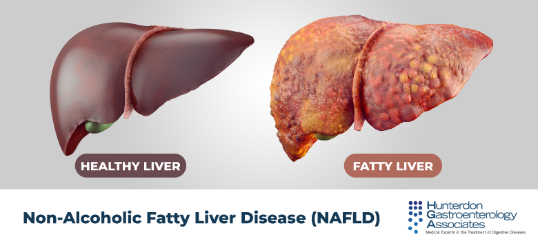
Nonalcoholic Fatty Liver Disease
Dyslipidemia is common among persons with NAFLD and plays a critical role in the development of cardiovascular disease, the leading cause of mortality among these persons. Dyslipidemia associated with NAFLD is typically characterized by a pattern of hyper-triglyceridemia, increased LDL levels, and reduced high-density lipoprotein (HDL) levels. Although patients with NAFLD have a higher risk of cardiovascular complications compared with the general population, NAFLD and nonalcoholic steatohepatitis (NASH) are not considered traditional risk factors for cardiovascular disease. Therefore, cardiovascular risk should be assessed according to the 2013 American College of Cardiology/American Heart Association guidelines, which use presence of known coronary artery disease, cardiovascular risk factors, and 10-year cardiovascular event risk assessment to determine the need for treatment with lipid-lowering agents and to establish LDL targets for primary and secondary cardiovascular risk prevention (Table 2; discussed later).
Diet and exercise are the first-line approaches to reduce LDL levels. When diet and exercise are insufficient, statins are the first-line pharmacologic agents for LDL reduction and have proven benefit for primary and secondary prevention of cardiovascular disease. Several studies have demonstrated that statins are effective in reducing LDL levels and cardiovascular events and are safe in patients with NAFLD. Intensive statin therapy (eg, pravastatin, 80 mg daily) was associated with a significant reduction in LDL levels compared with placebo among patients with chronic liver disease primarily caused by NAFLD (mean percentage LDL reduction, 30.6%; standard deviation, 16.5%).10 In a post hoc analysis of the ATTEMPT study, a greater benefit in primary cardiovascular disease prevention was observed among patients with NAFLD and metabolic syndrome in whom LDL reduction was aggressive with intensive statin therapy (atorvastatin titrated from 10 to 80 mg daily) aimed to reduce LDL levels to <100 mg/dL compared with those in whom the LDL reduction was aimed to reduce the level to <130 mg/dL.11 Post hoc analyses of 2 large randomized controlled trials (RCTs) (GREACE and IDEAL studies) also found superior secondary cardiovascular disease prevention benefit in association with intensive statin therapy among patients with increased aminotransferase levels attributed to NAFLD. In the IDEAL study, compared with moderate statin therapy, intensive statin therapy (atorvastatin, 80 mg daily) was associated with a 44% relative risk reduction in secondary cardiovascular events (hazard ratio, 0.556; 95% confidence interval, 0.367–0.842).12 In the GREACE study, compared with usual care, intensive statin therapy (atorvastatin titrated from 10 to 80 mg daily for an LDL goal of <100 mg/dL) was associated with a 68% relative risk reduction (P < .0001).11 Both studies found greater cardiovascular benefit among patients with increased aminotransferase levels than in those with normal levels.
Statins are well tolerated and not associated with liver-related adverse events among patients with NAFLD and dyslipidemia.10,13,14 In RCTs, patients with NAFLD who were treated with intensive statin therapy experienced significant reductions in aminotransferase levels compared with control subjects.10 Observational studies have similarly reported significant reductions in amino-transferase levels and steatosis on liver biopsy specimens in statin-treated patients with NAFLD. However, histologic improvements in ballooning and fibrosis have not been observed in association with statins.
Experience with ezetimibe in patients with NAFLD is much less extensive than that with statins. Treatment with ezetimibe, 10 mg daily, was associated with significant reductions in LDL and total cholesterol levels among patients with NAFLD in several observational studies15–18 and 1 small open-label RCT.19 However, there have been no studies looking at the effect of ezetimibe on cardiovascular disease prevention in patients with NAFLD. Ezetimibe may be associated with improvements in aminotransferase levels. Mixed results from studies evaluating the effect of ezetimibe on liver histology in patients with NASH suggest that ezetimibe may improve steatosis, NAFLD activity score, and hepatocyte ballooning but not fibrosis.15,17,19–23
Omega-3 fatty acids and fenofibrates are safe and effective for the treatment of isolated hyper-triglyceridemia. Neither medication is associated with improvement in liver histology in patients with NAFLD.
In summary, statin therapy is indicated for treatment of increased LDL levels among patients with NAFLD who are at increased risk for adverse outcomes of cardiovascular disease. Statin therapy is associated with reductions in LDL levels and prevention of cardiovascular disease. Ezetimibe may also be used for treatment of increased LDL levels and is associated with reductions in these levels, but its efficacy for cardiovascular disease prevention is unknown. Omega-3 fatty acids and fibrates are indicated for the treatment of hypertriglyceridemia. All of these agents are safe and well tolerated in patients with NAFLD. However, there is no conclusive evidence to date that treatment of dyslipidemia with any agent (statins, fibrates, omega-3 fatty acids) improves histology or liver-related outcomes in patients with NASH.
Viral Hepatitis
Hepatitis C
Hepatitis C virus (HCV) chronically infects up to 170 million individuals globally and can lead to the development of cirrhosis, hepatocellular carcinoma, and the need for liver transplantation.24 HCV replication impacts host lipid metabolism via several mechanisms. HCV virions interacts with circulating lipoproteins to infect hepatocytes via the LDL receptor and interacts with the cell surface markers Niemann-Pick C1-like 1, a receptor for cholesterol resorption, and scavenger receptor class B member 1, which promotes uptake of cholesterol from lipoproteins.25–27 Once inside the hepatocyte, HCV interacts with host cytosolic lipid droplets and diacylglycerol O-acetyltransferase 1 to form the HCV core protein and uses an intermediate of the cholesterol synthesis pathway for replication.27 HCV also uses host lipids for secretion by complexing with apoE-containing very-low-density lipoproteins and HDL.28
These HCV-host interactions affect host circulating lipid levels. Both acute and chronic HCV infections lead to a decrease in serum LDL and total cholesterol levels.29,30 However, the lower lipid levels found in persons with HCV infection do not confer a decreased risk of cardiovascular disease; on the contrary, chronic HCV infection is associated with an increased risk of acute myocardial infarction when compared with matched control subjects.31,32 Furthermore, LDL and total cholesterol levels rebound after spontaneous and treatment-induced viral clearance. Therefore, in patients with HCV infection, lipid levels should be monitored, with a special focus on lipid levels following HCV clearance, to determine if treatment of dyslipidemia is needed. Treatment with statins is safe in individuals with liver disease, including hepatitis C, and should be first-line therapy.33
Hepatitis B
Hepatitis B virus (HBV) chronically infects more than 240 million individuals worldwide and can lead to end-stage liver disease, hepatocellular carcinoma, and the need for liver transplantation.33,34 Like HCV, HBV interacts with lipid metabolism in the host. HBV uses the peptide Naþ-taurocholate cotransporting polypeptide as a point of viral entry and an aid in the production of hepatitis B surface antigen. The binding of HBV to Na+ taurocholate cotransporting polypeptide impairs bile acid uptake, thereby leading to increased bile acid synthesis and bile acid conversion to cholesterol.35 Hepatitis B surface antigen formation also relies, in part, on the host endoplasmic reticulum lipid bilayer and, once formed, is exported as a lipoprotein particle.36
Expression of lipogenic genes may also be altered by HBV infection. Mouse models of HBV infection demonstrate increased expression of genes for SREBP2, 3-hydroxy-3-methylglutaryl-coenzyme A reductase, the LDL receptor, fatty acid synthase, ATP citrate lyase, peroxisome proliferator-activated receptor-ɣ, and apolipoprotein A1.37,38
Data on host lipid levels during HBV infection in humans are limited. A case-control study found that HBV-infected individuals had lower serum triglyceride and HDL levels than age-matched control subjects. A second retrospective study found that serum HBV DNA levels were inversely correlated with serum triglyceride levels. No relationship between serum HDL and HBV DNA levels was seen.39
As for HCV infection, there is a lack of data to guide lipid management in individuals with HBV infection. Clinicians should adhere to guidelines from the American College of Cardiology/American Heart Association on lipid management (Table 2).1 Statin therapy is safe to use in patients with chronic hepatitis B and should not be withheld.
Primary Biliary Cholangitis
Primary biliary cholangitis (PBC) is a chronic infiammatory autoimmune cholestatic liver disease associated with dyslipidemia. Individuals with PBC have a complex pattern of dyslipidemia (reviewed in Ref.40). Patients with PBC have increased serum triglyceride levels and variable HDL levels: patients with PBC and stage 1 or 2 fibrosis have increased HDL levels, those with stage 3 fibrosis have variable HDL levels, and those with stage 4 fibrosis have decreased HDL levels. These patients have increased total cholesterol levels largely because of elevations of lipoprotein X. Lipoprotein X is antiatherogenic; therefore, even though patients with PBC have dyslipidemia, the dyslipidemia may not lead to an increase in cardiovascular events. It is estimated, however, that about 10% of patients with PBC have a significant risk of cardiovascular disease and should be treated with medications to reduce that risk. Statins, ezetimibe, and fibrates have all been shown to be safe in patients with PBC.
Fibrates are second-line therapies for the treatment of cholestasis in patients with PBC and also affect serum lipids.41 Fibrates have anticholestatic effects through the activation of peroxisome proliferator-activated receptor and downregulation of bile acid synthesis pathways. Benzafibrate, 400 mg daily, alone or in combination with ursodeoxycholic acid, has been shown to lower serum alkaline phosphatase levels in small studies, but concern remains about ascertainment bias and the significance of biochemical rather than histologic or survival endpoints (reviewed in Ref.41). A large RCT is underway, and until the results are available, these medications may be used off-label in patients in whom the response to ursodeoxycholic acid is inadequate. Fibrates, however, do not significantly reduce the risk of cardiovascular events42 and should not be used for primary prevention of cardiovascular disease in patients with PBC and dyslipidemia.
Obeticholic acid (OCA) is a second-line therapy for the treatment of PBC and also affects serum lipids.41 OCA is a farsenoid-X receptor agonist that directly regulates genes involved in bile acid synthesis, secretion, transport, absorption, and detoxification. The phase 3 PBC OCA International Study of Efficacy (POISE)43 showed a 33%–39% reduction in serum alkaline phosphatase levels in the group receiving 10 mg daily compared with 5% in the group receiving placebo (P < .001) at 12 months of treatment. However, patients taking OCA had an increase in LDL and total cholesterol levels and a decrease in HDL levels, the cardiovascular consequences of which remain to be determined. If cardiovascular risk is indeed increased in patients taking OCA, the medication may need to be coupled with a second medication to lower that risk. Thus, until more data are available, OCA should be avoided in patients with cardiovascular disease or risk factors for cardiovascular disease. Additionally, the recommended initial dose for patients with moderate to severe liver impairment (Child-Pugh B and C) is 5 mg once weekly, rather than the 5 mg daily used for other PBC patients (https://www.fda.gov/downloads/Drugs/DrugSafety/UCM576880.pdf). When daily dosing has been administered inadvertently in Child-Pugh B and C patients, serious liver injury and death have been reported. Furthermore, up to 10% of patients discontinued treatment because of pruritus.
Several studies have now shown that statins can lower LDL levels in patients with PBC who also have increased rates of cardiovascular disease. Atorvastatin, 10 mg daily for 1 year, in early stage PBC reduces total cholesterol, LDL cholesterol, LDL, and triglyceride levels without affecting progression of cholestasis.44 Long-term atorvastatin use also led to reductions in total and LDL cholesterol levels.45 Similar reductions in total cholesterol and LDL cholesterol levels were seen in patients treated with simvastatin, 20 mg daily.46 Although initially considered promising treatments for PBC,46 recent trials have not shown an effect of statins in reducing serum alkaline phosphatase levels.44,45 Studies also show that ezetimibe is safe in patients with PBC.47 Therefore, statins and ezetimibe should be considered in patients with PBC with cardiovascular disease risk factors.
Cirrhosis
Although cirrhosis was previously thought to protect against atherosclerotic disease, work in recent years has demonstrated that the prevalence of coronary artery disease among patients with cirrhosis may be higher than that in the general population.48–50 Cardiovascular risk varies according to etiology of liver disease and is highest in cirrhosis caused by alcohol, HCV, and NASH51–53; in NASH, the risk is mediated by concomitant risk factors for cardiovascular disease or by the presence of steatosis and insulin resistance. The risk of atherosclerosis is highest in NASH cirrhosis.53 Cholestatic liver diseases, however, do not carry an overall increased risk of atherosclerotic disease.51
Statins are safe and can be used in patients with Child-Pugh class A cirrhosis.9 There are little to no available data regarding the safety and risks of statin use in patients with decompensated cirrhosis. The updated 2014 recommendations of the Liver Expert Panel assembled to address the safety of statins in liver disease9 provides some guidance and advises against statin use among patients with Child-Pugh class B or C cirrhosis. The underlying rationale is that the generally grave liver-related prognosis of patients with Child-Pugh class B or C cirrhosis makes it unlikely that they will benefit from the cardiovascular benefits associated with lipid-lowering therapy. In addition, moderate-to-severe hepatic impairment may result in reduced drug metabolism and consequently abnormally high serum drug levels (although an increased risk of hepatotoxicity has not been demonstrated).
In addition to lipid-lowering effects, statins are associated with reductions in portal pressure and may reduce the risk of decompensation among patients with HBV or HCV cirrhosis.54–56 However, the data are still evolving, and use of statins to reduce portal pressure is not the standard of care. Therefore, statins should not be used for the treatment of portal hypertension.
Post-Transplant Dyslipidemia
Dyslipidemia is common following liver transplantation, occurring in up to 62% of transplant recipients.57 Post-transplant dyslipidemia is seen most commonly in patients with obesity or diabetes mellitus pretransplantation but can develop in the absence of these comorbid conditions. Weight gain post-transplantation and use of immunosuppressant medications, including calcineurin inhibitors and the mechanistic target of rapamycin inhibitor sirolimus, also increase the risk of post-transplant dyslipidemia.58
Lipid-lowering agents, specifically statins, are safe in the post-transplant population and should be used as needed to treat dyslipidemia.59 Calcineurin inhibitors and several statins are metabolized by cytochrome P-450 3A4, and concurrent use may increase the risk of statin-associated myopathy. Pravastatin and fiuvastatin are not metabolized by cytochrome P-450 3A4 and, when used with cyclosporine, may not increase the risk of statin-associated myopathy.59,60
Conclusions
Lipid-lowering medications are safe and effective in lowering the risk of cardiovascular disease in individuals with compensated liver disease. Some lipid-lowering agents may help in the primary or secondary treatment of specific types of liver disease. Interactions with other drugs should be considered when choosing particular lipid-lowering medications. Only in patients with decompensated cirrhosis and in those with well-documented DILI from a lipid-lowering agent should these medications not be used.